Figure 7. Intravitreal injection of dorsomorphin aggravated retinal damage. A, B: Statistical analysis of retinal hematoxylin and eosin staining and retinal ganglion cell quantity in mice treated with dorsomorphin
(7 μM, 2 h) for 1 and 3 days shown. These retinas have not received treatment for high intraocular pressure. *Significantly
different from control. ***p < 0.001. #Significantly different from dorsomorphin. ###p < 0.001, significantly different from dorsomorphin for 1 day. aaap < 0.001. n = 6. Scale bar: 50 μm.
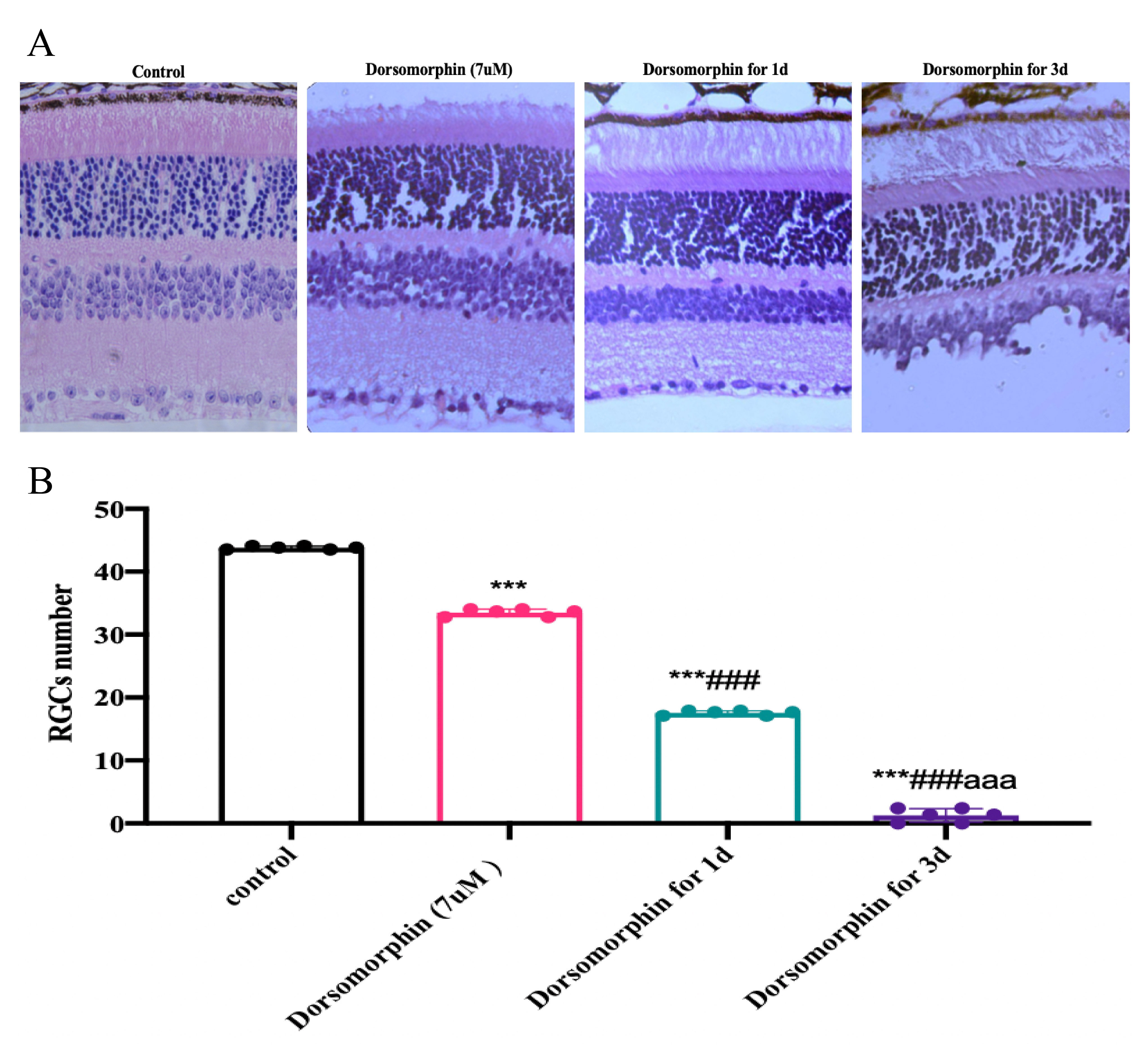
 Figure 7 of
Cui, Mol Vis 2026; 32:265-279.
Figure 7 of
Cui, Mol Vis 2026; 32:265-279.