Figure 7. Ultrastructural and molecular characterization of hIPSC-derived photoreceptor-like cells. A: Schematic representation of a photoreceptor (PhR) cell highlighting key subcellular structures: discs, synapses, nuclei
(Nu), endoplasmic reticulum (ER), mitochondria (m), Golgi apparatus (g), opsin-carrying vesicles (OCV), calyceal processes,
outer segment (OS), inner segment (IS), connecting cilia (CC), and microtubules (mt). Transmission electron microscopy (TEM)
of hIPSC-derived PhR-like cells at day 50 of differentiation (D50, Exp 3) reveals nuclei (Nu), IS, OS, calyceal processes
(arrow), mitochondria (m), and putative OCVs. Scale bar: 10 μm. B: Heat map showing expression of genes associated with the connecting cilium across three differentiation experiments. Data
are presented as gene-wise scaled z-scores. C: Heat map showing expression of genes associated with photoreceptor outer segments across three differentiation experiments.
Data are presented as gene-wise scaled z-scores. D: TEM image of hIPSC-derived PhR-like cells at day 106 of differentiation (D106, Exp 3) showing abnormal organization of putative
outer segments, appearing as ‘whorls’ (highlighted in yellow box). Scale bar: 5 μm. E: Immunofluorescence analysis of RHO, OPSN-S, CX36, and TMEM138 expression at day 111 of differentiation (D111). Nuclei were
counterstained with Hoechst 33342.
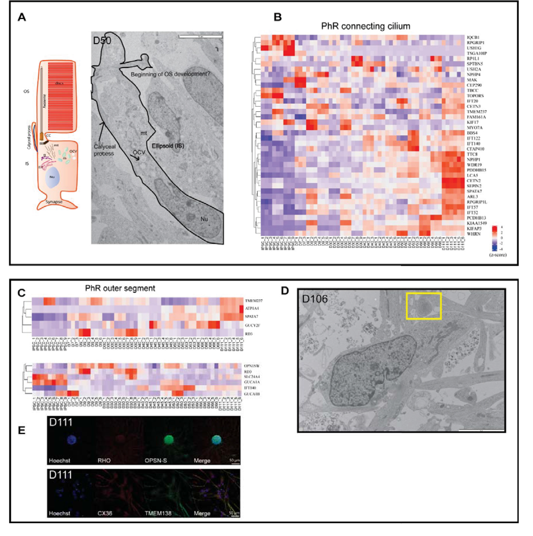
 Figure 7 of
Beaver, Mol Vis 2026; 32:232-xxx.
Figure 7 of
Beaver, Mol Vis 2026; 32:232-xxx.