Figure 5. Flow cytometric analysis of retinal marker expression during differentiation. A. Flow cytometry of cells harvested from Experiment 2 at D52 showed 97.4% RCVRN-mCherry–positive cells, comparable to 99.9%
positivity in 661W control cells. B. Analysis for Experiment 3 at D42 revealed 93.6% RCVRN, 96.2% OPSN-M/L, and 70.3% OPSN-S expression, relative to 661W positive
controls. At D49, 93.3% of the population was RCVRN-positive, compared to 99.3% in 661W cells.
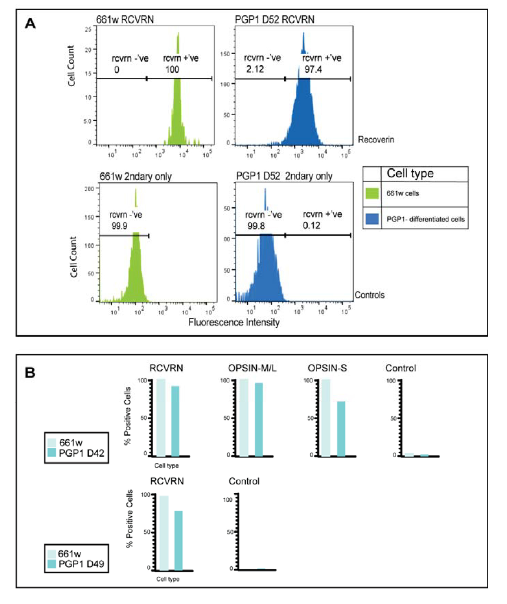
 Figure 5 of
Beaver, Mol Vis 2026; 32:232-xxx.
Figure 5 of
Beaver, Mol Vis 2026; 32:232-xxx.