Figure 4. Representative immunofluorescence images from two independent experiments showing retinal marker expression during late differentiation
(Days 52–66). A: At D52, cells expressed RCVRN, ARR3, CRX, OPSN-S, TUJ1, LHX2, and the proliferation marker Ki-67. B: At the same time point, NRL and RCVRN were absent, while THRβ, ARR3, OPSN-M/L were apparent. C: By D61, PRDM1 and CRX apeared absent, whereas ARR3, OPSN-S, GNAT2, RCVRN, and THRβ remained expressed. D: At D66, TUJ1 and LHX2 expression declined consistently across replicates, while RCVRN, OPSN-M/L, ARR3, OTX2, and OPSN-S
were detected; CRX was barely detectable. Cell nuclei were counterstained with Hoechst 33,342. Circular or clustered staining
patterns reflect regions of natural cell aggregation that occur at later differentiation stages due to preferential cell-cell
association within the culture.
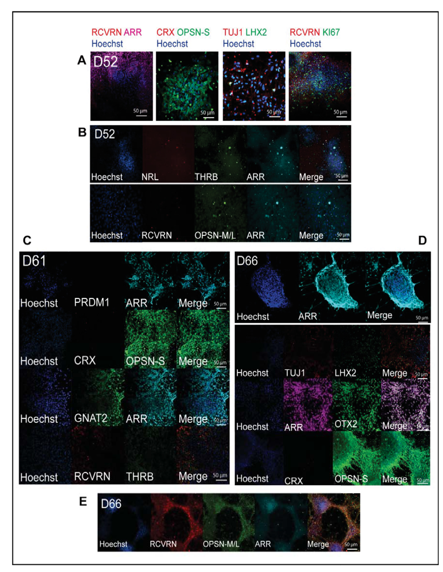
 Figure 4 of
Beaver, Mol Vis 2026; 32:232-xxx.
Figure 4 of
Beaver, Mol Vis 2026; 32:232-xxx.