Figure 9. Cellular signaling mediating the reducing effect of TGF-β1 on the TLR9 expression in cultured RPE cells. The TLR9 gene expression was determined with qPCR in RPE cells cultured in absence or presence of hypoxia mimetic CoCl2 (150 µM), respectively (A, B), in a 0.1% O2 atmosphere (C) or in presence of TGF-β1 (10 ng/ml; D) for 24 h. The RPE cells were pre-incubated with the inhibitors for 30 min. The following inhibitors were used: the inhibitor
of TGF-β1 superfamily activin receptor-like kinase receptors, SB431542 (10 µM), an anti TGF-β pan specific neutralizing antibody
(10 µg/ml), the Smad3 inhibitor, SIS3 (10 µM), the CREB inhibitor 666–15 (250 nM), the ERK1/2 inhibitor, PD98059 (20 µM),
the p38 MAPK inhibitor, SB203580 (10 µM), the JNK inhibitor SP600125 (10 µM), the inhibitor of PI3K-kinases, LY294002 (5 µM).
DMSO (1:1000) served as solvent for the inhibitors. The data are displayed as fold change of unstimulated control (1). ACTB and B2M were used to normalize the results. Each bar shows data obtained from 3 to 6 independent experiments with cells from different
donors. One sample t test was used for statistical analysis. Significant difference versus unstimulated control: *p<0.05; **p<0.01. Significant
differences versus CoCl2 control (B), versus 0.1% O2 (C) or versus TGF-β1 control (D): ●p<0.05. .
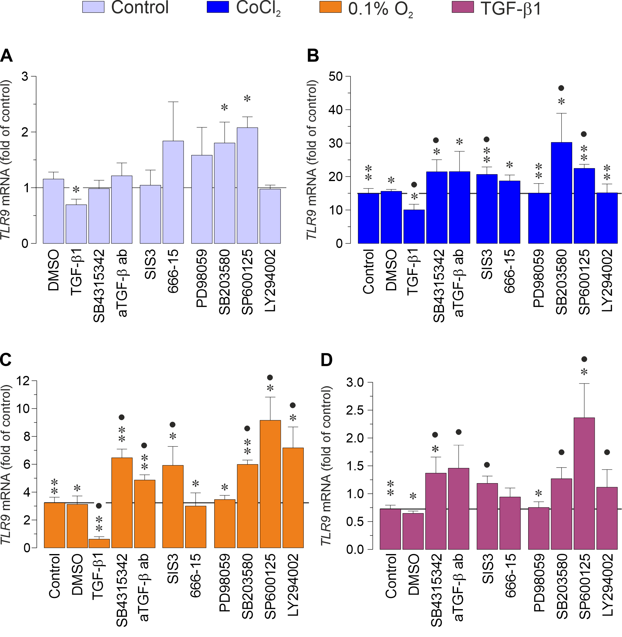
 Figure 9 of
Esser, Mol Vis 2026; 32:210-230.
Figure 9 of
Esser, Mol Vis 2026; 32:210-230.