Figure 7. Effects of the TLR9 inhibiting oligonucleotide iODN INH-18 on the activation of NF-κB under chemical hypoxia (150 µM CoCl2) or 0.1% O2 atmosphere in human RPE cells TLR9 was blocked with iODN INH-18 (2.5 µM). The cells were cultured for 24 h in normoxic or
hypoxic conditions (CoCl2, 0.1% O2 atmosphere) as indicated by the panels of the bars. The level of total proteins and phosphorylated proteins were determined
with western blotting. As negative control co-ODN 2088 (co-ODN; 2.5 µM) was used. A, B: Representative western blots of different total and phosphorylated signaling proteins using 25 µg whole cell protein extract
for total protein and 35 µg for phosphorylated proteins. β-actin was used as loading control. The image shows one example
of 3 experiments. C. Graphical illustration of western blotting results of phospho-NF-κB p65 (pNF-κB p65) are presented. Signals quantified by
densitometry and normalized with β-actin are shown in relation to the untreated control (100). Each bar represents data from
3 independent experiments with cells from different donors. One-way ANOVA followed by Bonferroni’s multiple comparison test
was used to analyze the results Significant difference versus. unstimulated control: *p<0.05. Significant differences versus CoCl2 or 0.1% O2 controls: ●p<0.05. .
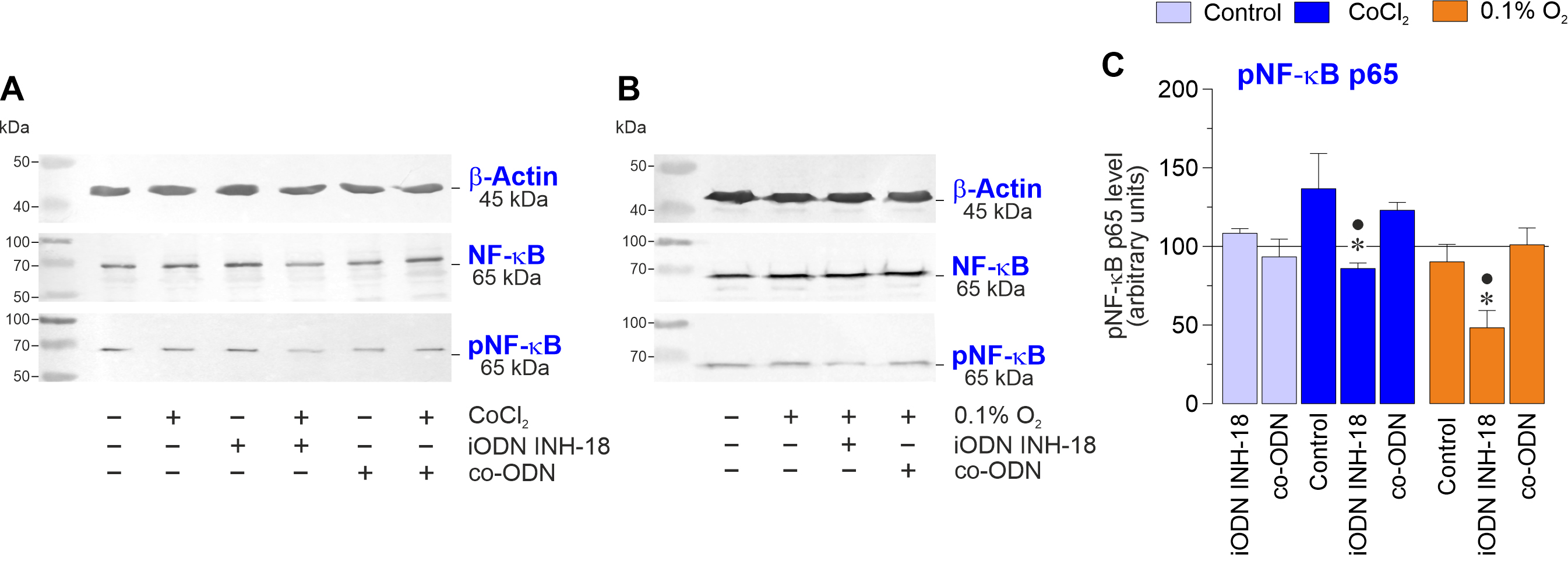
 Figure 7 of
Esser, Mol Vis 2026; 32:210-230.
Figure 7 of
Esser, Mol Vis 2026; 32:210-230.