Figure 6. Effects of inhibitory oligonucleotides for TLR9 on the hypoxic gene and protein expression in cultured human RPE cells. The
cells were stimulated for 24 h with CoCl2 (150 µM) or in 0.1% O2 atmosphere as indicated by the panels of the bars. TLR9 was blocked by iODN 2088 (1 µM) or iODN INH-18 (2.5 µM). As negative
control, control co-ODN 2088 (co-ODN; 1 and 2.5 µM) was used. Results are shown as fold change to unstimulated controls (1).
TGF-β1 protein secretion was analyzed in cell culture supernatants using ELISA. Results are shown as per cent to CoCl2 or to 0.1% O2 controls (100). The gene expression of A: TLR9B: TLR9 adaptor protein MYD88. C: Transcription factor IRF7. D: Transcription factor RELA (the gene of p65 subunit of transcription factor NF-κB). E: IFNA, F: COX2, G: MMP9. The data are shown as fold change of unstimulated control (1). ACTB and B2M were used to normalize the results. H: Shows total TGF-β1 protein amounts secreted by RPE cells cultured in presence of CoCl2 or a 0.1% O2 atmosphere. Each bar represents data (mean ± SEM) obtained in 4–7 (iODN 2088) or 3–4 (iODN INH-18) independent experiments
using cells from different donors. Mann–Whitney-U-Test was used for statistical analysis. Significant difference versus unstimulated
control: *p<0.05, **p<0.01. Significant differences versus CoCl2 or O.1% O2 controls: ●p<0.05. .
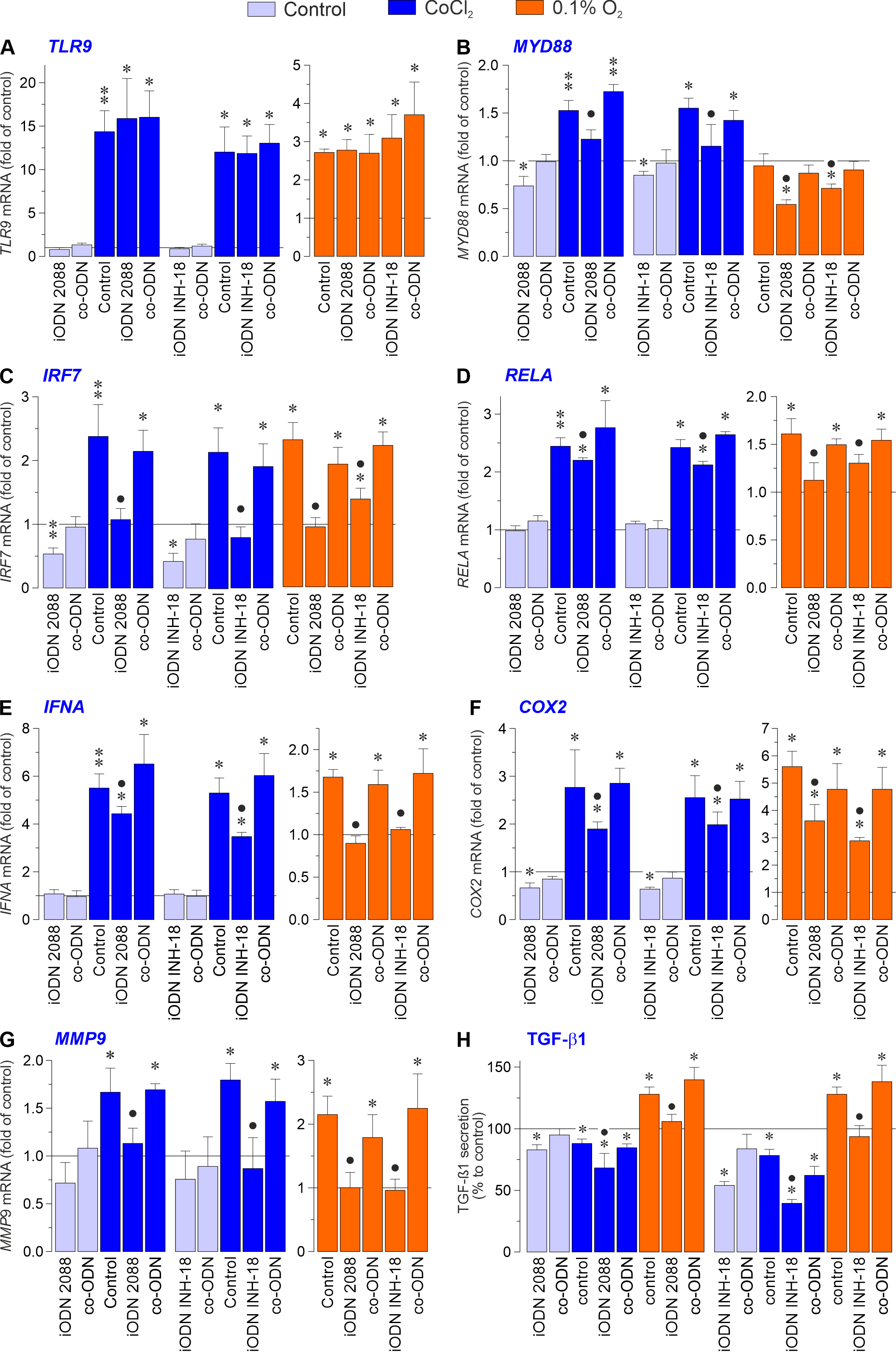
 Figure 6 of
Esser, Mol Vis 2026; 32:210-230.
Figure 6 of
Esser, Mol Vis 2026; 32:210-230.