Figure 3. TLR9 protein in cultured human RPE cells. A: Immunolabeling of cultured RPE cells with an anti-TLR9 antibody (green) and an antibody against the tight junction protein zonula occludens-1 (ZO-1; red). Cell nuclei are labeled with DAPI (blue). Bars, 20 µm. B: western blots of TLR9 protein performed with lysates of acutely isolated neuroretinas and cytosolic and membrane protein
extracts of cultured RPE cells derived from two donors (1, 2). C: Representative western blots of TLR9 protein with lysates of RPE cells incubated in presence of NaCl (+100 mM), hypoxia
mimetic CoCl2 (150 µM), TGF-β1 (10 ng/ml), in a 0.1% O2 atmosphere or Triamcinolone acetonide (Triam; 50 µM) for 24 h. β-actin served as loading control. D: Densitometric evaluation of the western blot results. β-actin was used to normalize the results. The data were obtained
from 3 (TGF-β1) and 4 (NaCl, CoCl2, 0.1% O2, Triam) independent experiments using RPE cells from different donors. Significant difference versus unstimulated control:
*p<0.05. The statistical analysis was performed using the One-way ANOVA followed by Bonferroni’s multiple comparison test.
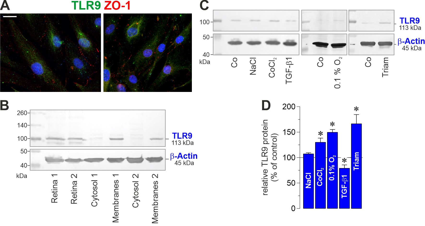
 Figure 3 of
Esser, Mol Vis 2026; 32:210-230.
Figure 3 of
Esser, Mol Vis 2026; 32:210-230.