Figure 5. Immunohisotochemical detection of-HIF-1α after 24 h incubation in 5 or 25 mM glucose. HIF-1α protein detection at time zero
and 24 h (B) exposed to 5 mM or 25 mM HIF-1α protein was detected at in the start of the incubation period in the explants irrespective
of glucose concentration. HIF-1α protein levels returned to undetectable basal levels in explants in 5 mM glucose at 24 h
but remained elevated in explants in 25 mM at 24 h (400X).
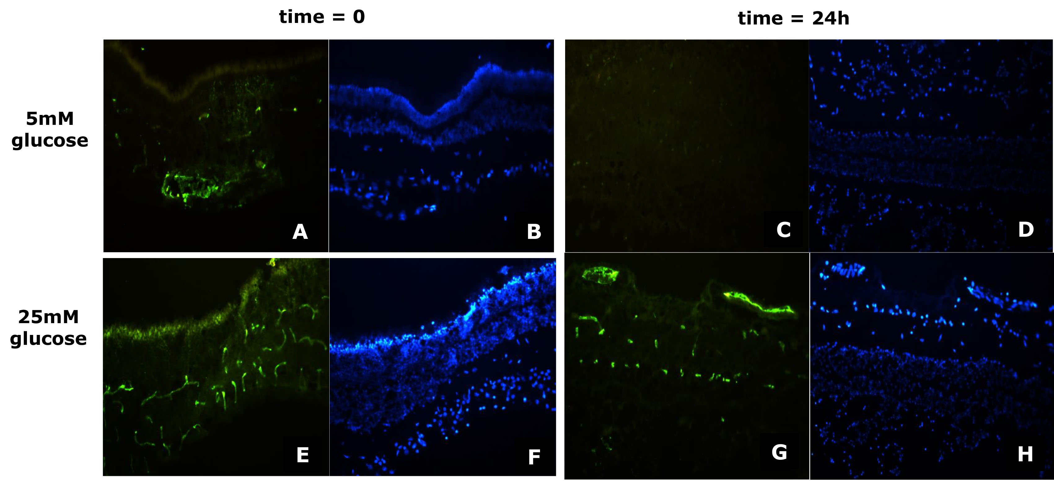
 Figure 5 of
Iwuagwu, Mol Vis 2026; 32:186-194.
Figure 5 of
Iwuagwu, Mol Vis 2026; 32:186-194.