Figure 2. Haematoxylin and eosin staining of retinal explants after 24 h incubation Selected micrographs of retinal explant at t=0 (40X)
and 24 h (20X) post culture. Isolated retinae cultured as explants have identifiable retinal layers; photoreceptors, outer
nuclear layer (ONL), outer plexiform layer (OPL), Inner nuclear layer (INL), Inner plexiform layer (IPL) and ganglion cell
layer (GCL).
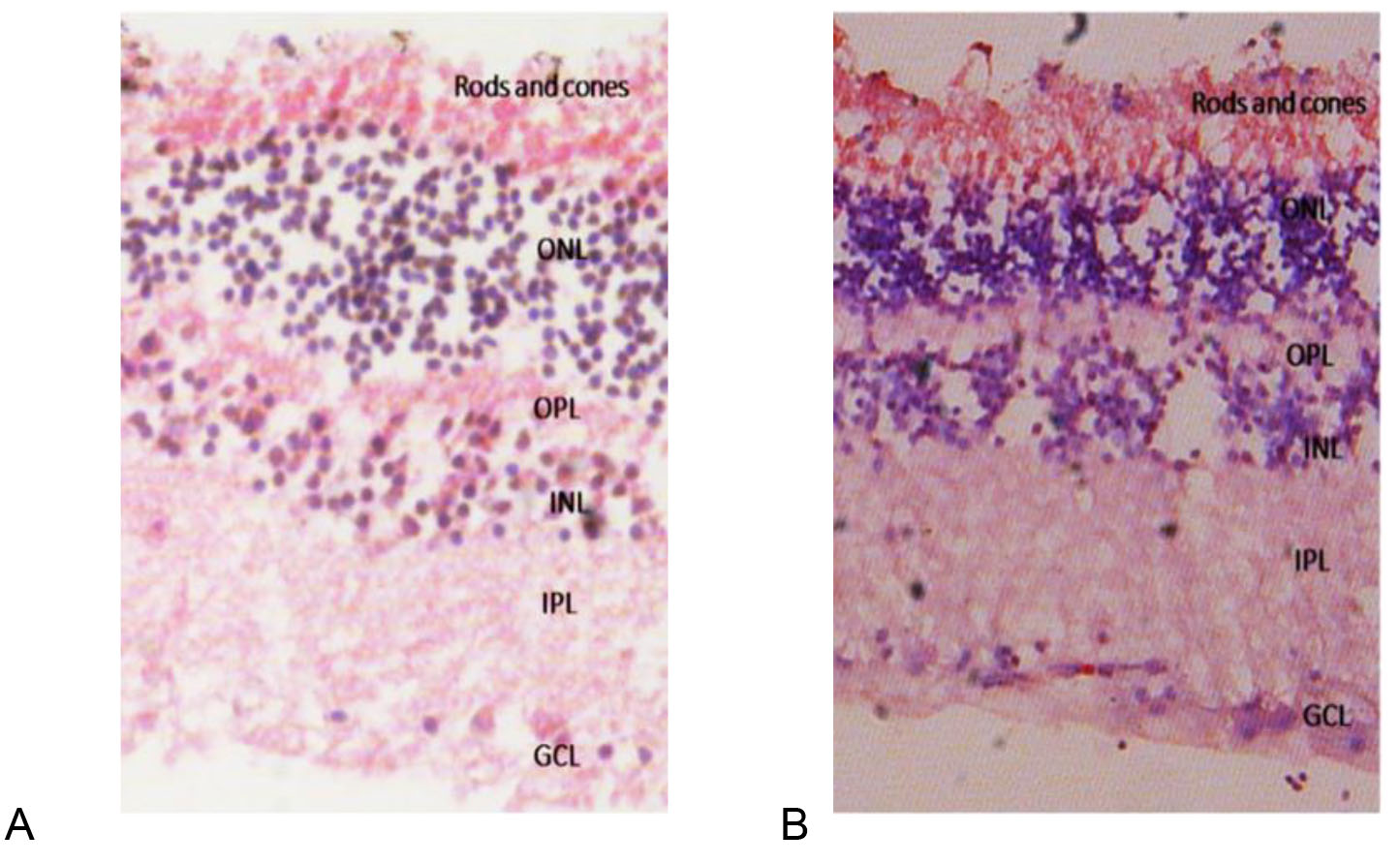
 Figure 2 of
Iwuagwu, Mol Vis 2026; 32:186-194.
Figure 2 of
Iwuagwu, Mol Vis 2026; 32:186-194.