Figure 1. Light microscopic images of explants and 2D control. Explant tissues and vessels showed vessel intactness and absence of degradation
for up to 72 h (A-D), there are signs of the beginning of tissue tear at 72 h (black arrowhead). Tissue and vessel degeneration was massively
evident after 24 h of culture in 2D plates and exaggerated with longer incubation (Black arrows; E-H).
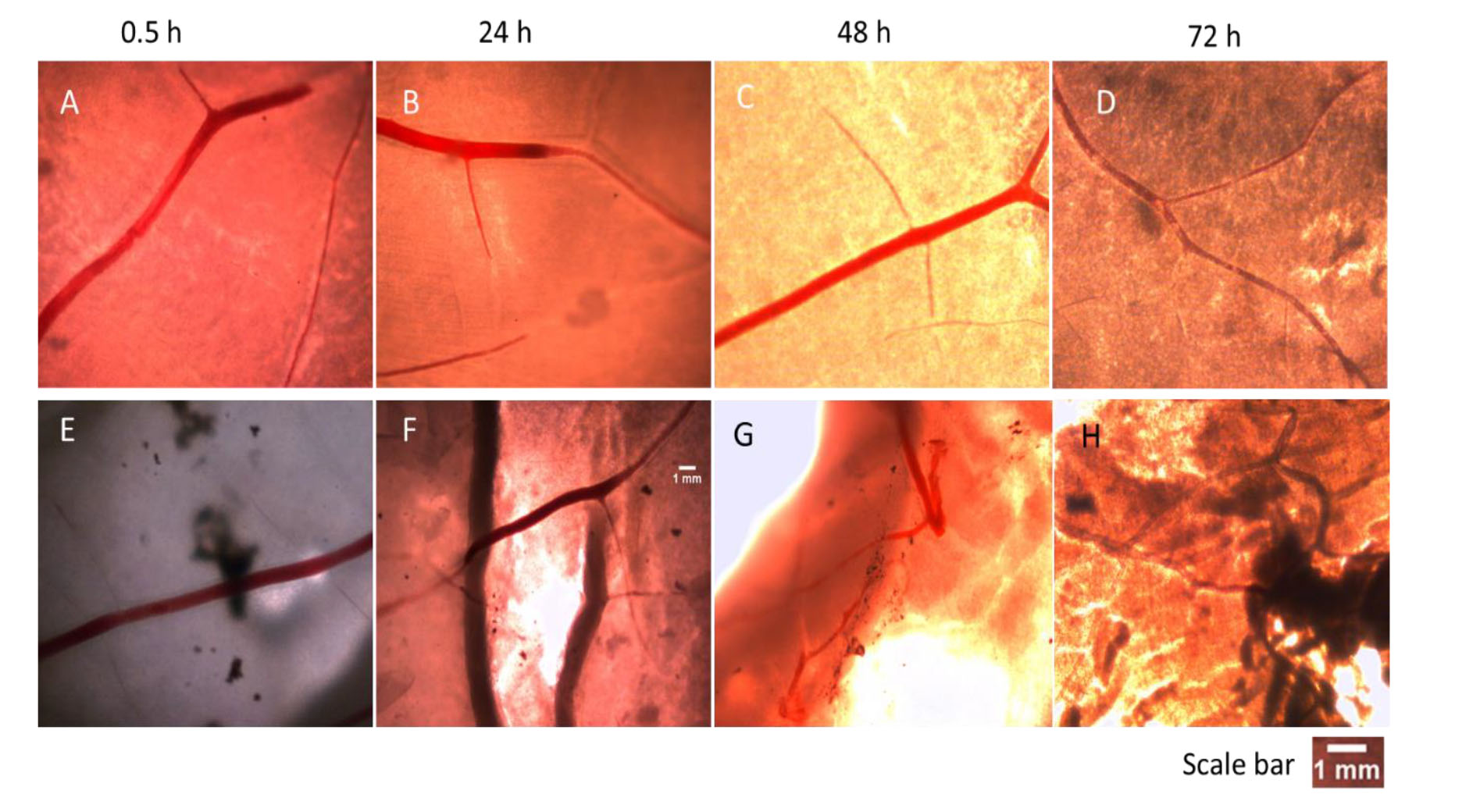
 Figure 1 of
Iwuagwu, Mol Vis 2026; 32:186-194.
Figure 1 of
Iwuagwu, Mol Vis 2026; 32:186-194.