Figure 8. Second harmonic generation (SHG) imaging results. Representative SHG images from control and WST11/NIR- treated rabbit scleral
specimens recorded from superficial (50 μm), mid (150 μm), and deep (300 μm) stroma. Crimped collagen fibril bundle architecture
does not display any marked differences between control and treatment groups.
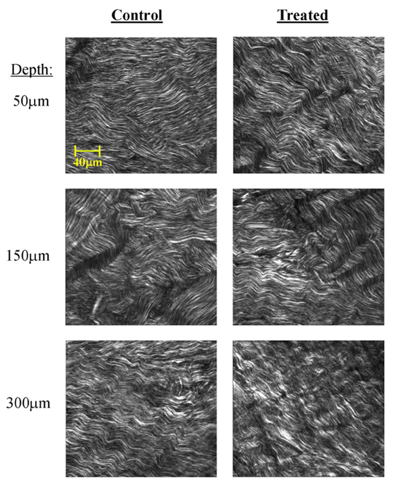
 Figure 8 of
Ma, Mol Vis 2026; 32:169-184.
Figure 8 of
Ma, Mol Vis 2026; 32:169-184.