Figure 10. Overlay of two-photon fluorescence (TPF) and second harmonic generation (SHG) images of sclera in the WST11/NIR-treated group
at a depth of 50 μm. Arrowheads: similar crimped morphology between the treatment-induced wavy structures in TPF and the collagen
in SHG.
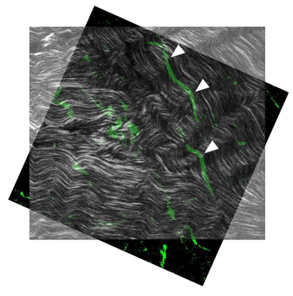
 Figure 10 of
Ma, Mol Vis 2026; 32:169-184.
Figure 10 of
Ma, Mol Vis 2026; 32:169-184.