Figure 1 of
Ma, Mol Vis 2026; 32:169-184.
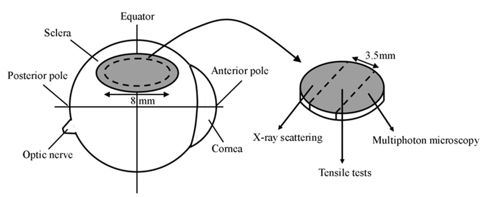
Figure 1.
Location of WST11/NIR treatment area (shaded region) on the rabbit eye and the use of dissected scleral specimens. Broken lines denote dissection cuts.
 Figure 1 of
Ma, Mol Vis 2026; 32:169-184.
Figure 1 of
Ma, Mol Vis 2026; 32:169-184.