Figure 2. Photomicrographs from stained retinas are shown for all six strains of mice used in this study: 129X1/SvJ, A/J, BALB/cByJ,
C57BL/6J, CBA/CaJ, and CAST/EiJ. The retinas stained for POU6F2 (red, A, E, I, M, Q and U) and BRN3A (green, B, F, J, N, R and V). The retinas were counter strained with TO-PRO-3 (blue, C, G, K, O, S and W) to label all of the nuclei in the retinal ganglion cell layer. Merged images of all three channels are shown (D, H, L, P, T and X). Notice that some strains have higher numbers of POU6F2 RGCs (BALB/cByJ) while other strains have relatively few POU6F2
RGCs (CAST/EiJ). Scale bar in X equals 50 µm.
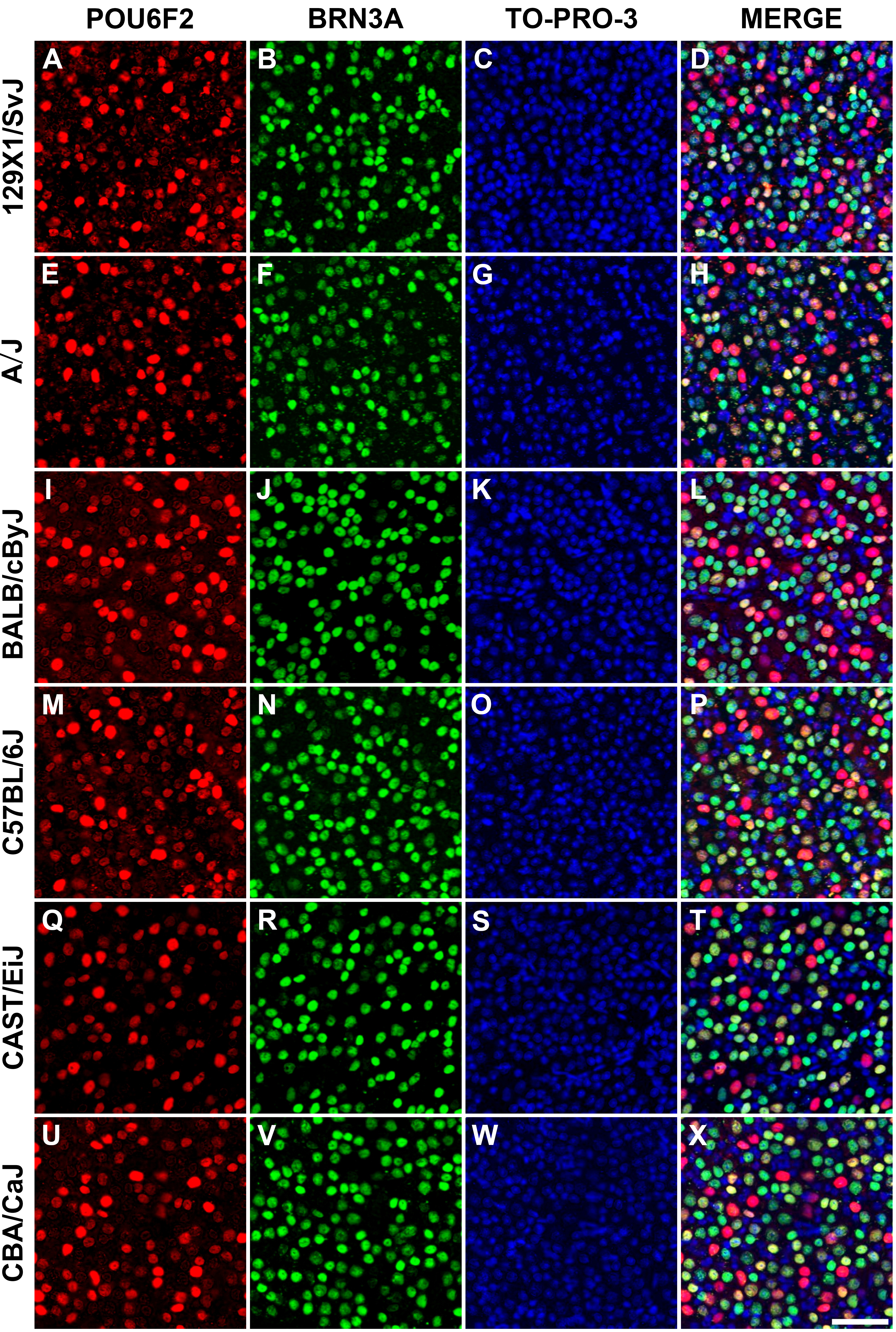
 Figure 2 of
Lin, Mol Vis 2026; 32:120-129.
Figure 2 of
Lin, Mol Vis 2026; 32:120-129.