Figure 3. Expression and safety of shEgr1 and mEgr1 transgenic elements in wt rods. A-C: Fundus images and OCT scans (top rows) and immunofluorescence images (bottom rows) of wt retinas subretinally injected with
(A) AAV::mOP-ctrl, (B) AAV::mOP-shEgr1 and (C) AAV::mOP-mEgr1 at 8 wpi. White arrows point to injection sites. OCT scans are color-coded in red when across the optic nerve, or blue when
across the injection site. Immunofluorescence panels show the transduction pattern of the virus (GFP), individual EGR1 and
rhodopsin (RHO) signals as gray-scale images and the colored merge. Scale bars: 20 μm. D: Egr1 mRNA levels in retinas of wt mice injected with mOP-ctrl (green) or mOP-mEgr1 (purple) at 2 wpi. Shown are individual data points and means ± SD n=6. Unpaired one-tailed t test, p value as indicated. E: Egr1 mRNA levels in transduced (red) and untransduced (blue) retinal areas of RhoP23H/+ mice injected with mOP-ctrl or mOP-shEgr1 at 12 wpi. Corresponding retinal areas of the same eye are connected by lines. n=3–4. Paired one-tailed t test, p value for mOP-ctrl: 0.4567, p value for mOP-shEgr1 as indicated. F: Left: spidergrams showing the mean and range of the ONL thickness of wt mice injected with mOP-ctrl, mOP-shEgr1 or mOP-mEgr1 at 8 wpi. Measurements were taken every 200 µm from the optic nerve head (0 µm). Right: quantification of the ONL thickness
of the nasal (transduced) retina after AAV injections. Each dot represents the average thickness at a particular distance
(every 200 µm from 400 µm to 2200 µm) from the optic nerve head. Shown are means ± SD. One-way ANOVA with Tukey’s multiple
comparisons test, p values as indicated. n=5–7. ONL: outer nuclear layer; INL: inner nuclear layer; GCL: ganglion cell layer.
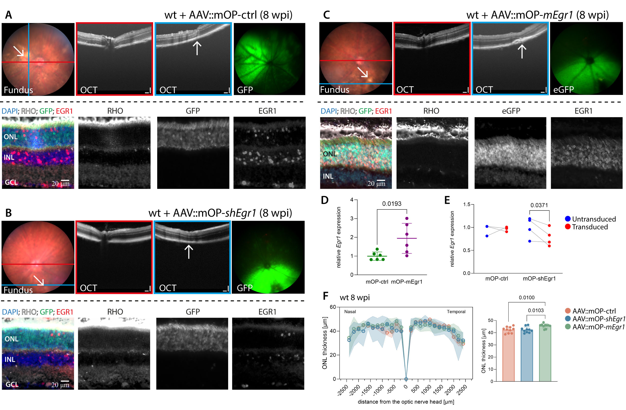
 Figure 3 of
Merolla, Mol Vis 2026; 32:102-118.
Figure 3 of
Merolla, Mol Vis 2026; 32:102-118.