Figure 2. AS-IV can improve the number of RGCs in model rats by promoting autophagy. A. Brn3a immunofluorescence staining was used to label RGCs in the retinal tissues of rats in all groups (n ≥ 3). B. Western blotting was used to assess LC3 protein levels in the retinal tissue of rats in all groups (n = 3). C. Real-time-PCR was used to assess the expression level of LC3 mRNA in the retina of rats in each group (n = 9). D. An immunofluorescence staining method was used to assess LC3 level in the retinal tissues of each group. E. TUNEL staining was used to label apoptotic cells (green) in the GCL (n ≥ 3); arrows point to apoptotic cells. Mean ± standard deviation; *p < 0.05 or **p < 0.01, compared with the MO group (excluding the CC group); #p < 0.05, comparison between the drug groups.
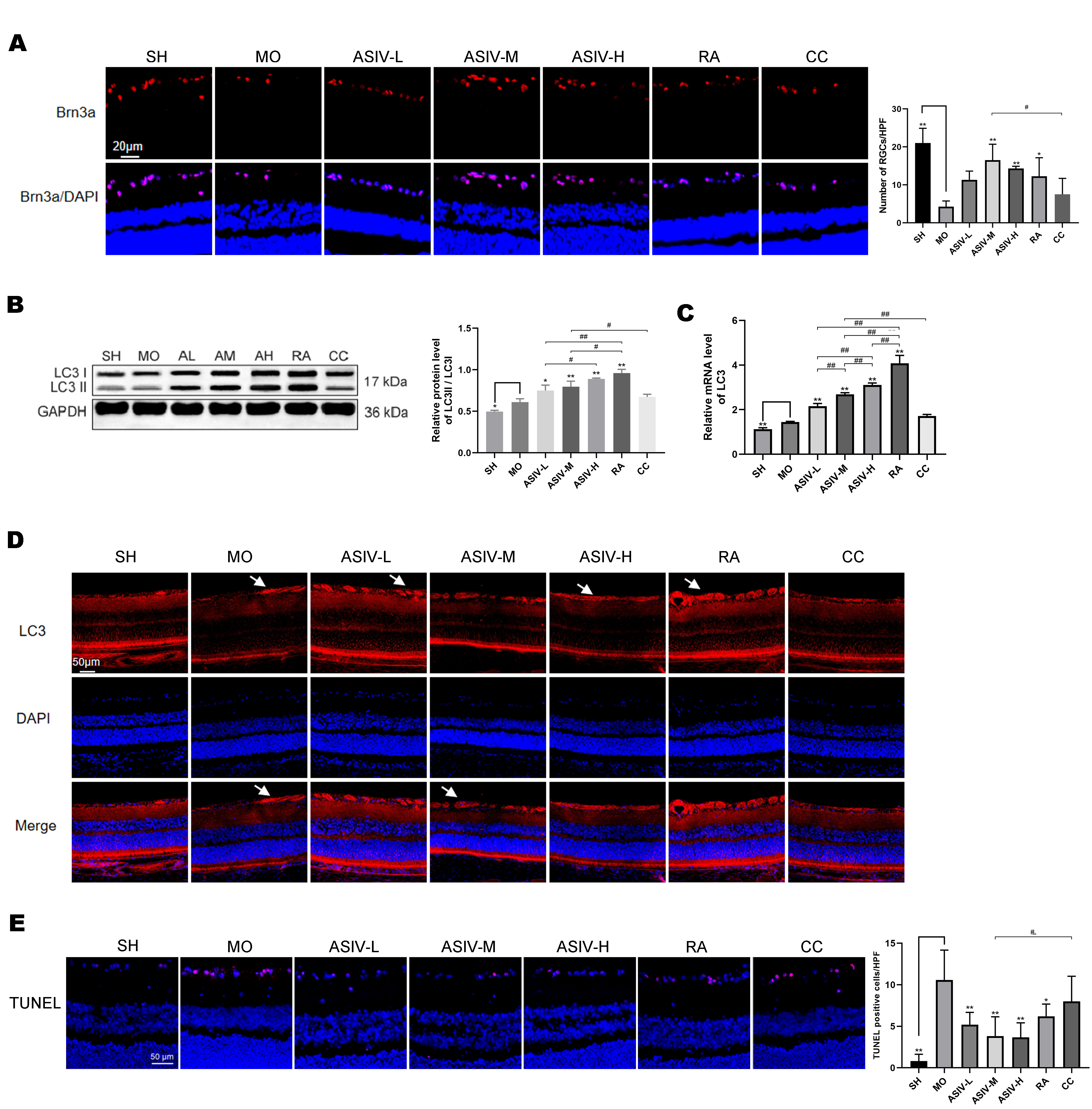
 Figure 2 of
Sun, Mol Vis 2025; 31:99-112.
Figure 2 of
Sun, Mol Vis 2025; 31:99-112.