Figure 7. The role of DNMT1 in cultured keratinocytes. Dnmt1F/F keratinocytes infected with Ad-GFP or Ad-Cre were examined for (A) Dnmt1 mRNA and (C) K14, K1 and K10 mRNA, normalized by mRNA of Gapdh. (B) DNMT1 protein normalized by β-actin. D: The percentage of apoptosis and proliferation quantified by immunolabelling with anti-Cas-3 and anti-EdU assays, respectively.
E: Immunofluorescent staining using anti-E-cadherin (E-Cad). Arrowhead points to Ad-GFP/Ad-GFP-Cre–infected cells. Scale bar:
30 µm. F: Dnmt1F/F reporter stable cells were infected with Ad-GFP or Ad-Cre, and luciferase activities were normalized by total protein. Values
are mean ± s.e.m. of three wells/adenovirus. *p < 0.05, ***p < 0.001 (two-tailed Student t test).
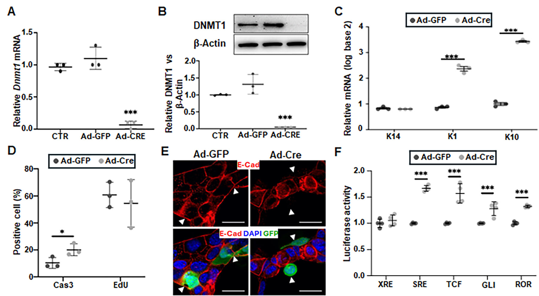
 Figure 7 of
Christianto, Mol Vis 2025; 31:85-97.
Figure 7 of
Christianto, Mol Vis 2025; 31:85-97.