Figure 6. Apoptosis and proliferation in wild-type and Dnmt1ΔOSE embryos. A: Representative images of the terminal deoxynucleotidyl transferase dUTP nick-end labeling assay showing apoptotic cells
(green, arrows) in different regions of the ocular surface epithelium. B: Illustration of the epithelial regions for the eyelids (red), cornea (green) and lens (yellow) used for quantification.
Quantification of (C) apoptotic cells and (D) proliferating cells in the different regions of the ocular surface epithelium in E14.5–E16.5 embryos. EL, Eyelid; CO, Cornea;
LN, Lens. Values are mean ± s.e.m. of three embryos/genotype. *p < 0.05, **p < 0.01 (two-tailed Student t test).
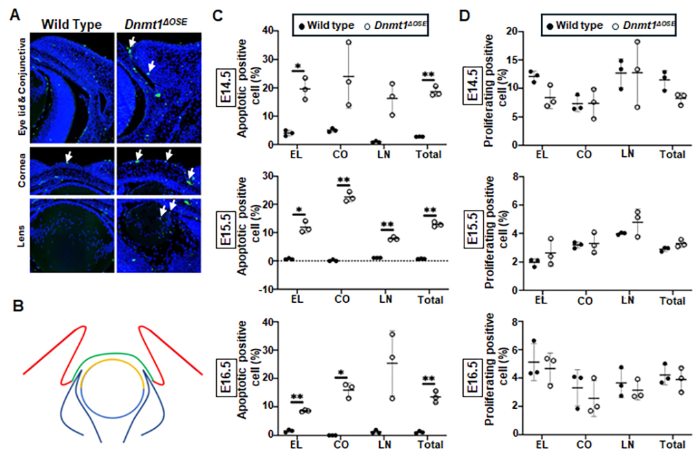
 Figure 6 of
Christianto, Mol Vis 2025; 31:85-97.
Figure 6 of
Christianto, Mol Vis 2025; 31:85-97.