Figure 5. DNA methyltransferase 1 ablation disrupts eyelid morphology and differentiation. A: Wild-type and Dnmt1ΔOSE E15.5 embryonic eye sections were subjected to Hematoxylin and eosin staining, and eyelid tip images were captured at lower
(upper panels) and higher magnifications of the selected areas (a, lower panels). Arrowheads point to the epithelial protrusion
of the eyelid tip, asterisks mark eyelid epithelial folding and arrows indicate cells displaying condensed nuclei in the Dnmt1ΔOSE eyelids. B: Immunostaining using anti-E-Cad and anti-ZO-1, as indicated. Arrows point to abnormal ZO-1 localization and reduced E-Cad
in the Dnmt1ΔOSE eyelids. C: Wild-type and Dnmt1ΔOSE embryonic eyelids at embryonic days indicated were subjected to immunostaining with anti-K10 for epithelial terminal differentiation.
Images were taken at low magnification (upper panels) and high magnification of selected areas (a, lower panels). Arrowheads
point to different K10 expression between wild-type and Dnmt1ΔOSE eyelid tips. Dashed lines indicate the epithelial basement membrane. Images are representative of at least three mice/genotypes.
Scale bars: 10 µm for (A) and (B). Scale bars: 30 µm for (C). EP, epithelium; DE, dermis.
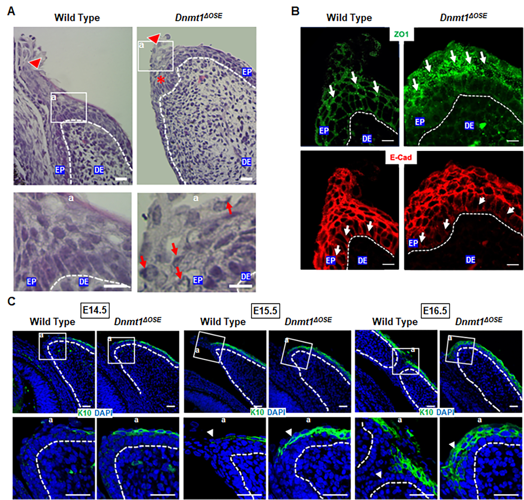
 Figure 5 of
Christianto, Mol Vis 2025; 31:85-97.
Figure 5 of
Christianto, Mol Vis 2025; 31:85-97.