Figure 3. DNA methyltransferase 1 maintains cornea integrity and differentiation. A: High magnification of hematoxylin and eosin-stained images of the corneas of wild-type and Dnmt1ΔOSE E16.5 embryos. The dashed lines indicate corneal stroma junctions with the epithelium and endothelium. Arrows point to abnormal-shaped
epithelial cells, asterisks show basal cells detached from the basement membrane and arrowheads point to the abnormal stromal
cells in the Dnmt1ΔOSE cornea. EP, epithelium, ST, stroma, ED, endothelium. B: Measurement of the stroma thickness (left) and quantification of keratocytes (right) in wild-type and Dnmt1ΔOSE corneas. Values are mean ± s.e.m. of three embryos/genotype. ***p < 0.001 (two-tailed Student t test). Immunohistochemistry of the corneas epithelium in E15.5 embryos using (C) anti-E-cadherin (E-Cad), (D) anti-ZO-1, (E) anti-K14 and (F) anti-K12 at low (upper panels) and high (lower panels) magnifications of the selected areas
(a, lower panels). Dotted lines indicate corneal epithelial basement membranes. White arrows indicate epithelial cell detachment
from the basement membrane. Images are representative of at least three mice/genotypes. Scale bars: 10 µm for (A), (C) and (D). 30 µm for (E) and (F).
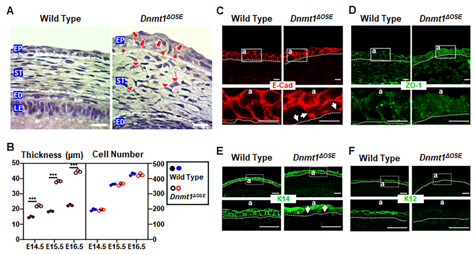
 Figure 3 of
Christianto, Mol Vis 2025; 31:85-97.
Figure 3 of
Christianto, Mol Vis 2025; 31:85-97.