Figure 4. Ocular status of posterior segment complications in in patient associated with LTBP2 variation. A: Anterior segment photograph showing a hypotonus eye with subluxated microspherophakic cataractous lens (marked by arrow)
in an eye with retinal detachment in case P9. B: B-scan ultrasound showing retinal detachment (arrow marked) in an eye post lensectomy in case P8. C: Shows B -scan with suprachoroidal haemorrhage (arrow marked) following combined trabeculotomy with trabeculectomy done elsewhere
in case P9.
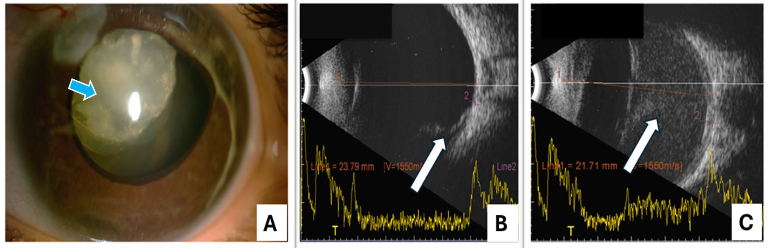
 Figure 4 of
Verma, Mol Vis 2025; 31:55-67.
Figure 4 of
Verma, Mol Vis 2025; 31:55-67.