Figure 3. Clinical status of eyes with secondary glaucoma, initially treated with lensectomy followed by glaucoma surgery as needed
in the study cohort. A: Advanced glaucomatous disc damage with posteriorly dislocated lens (marked by arrow) in case P3. B: Anterior segment picture of same eye of P3 after lensectomy. C, D: Anterior segment photograph of case P16 and case P7 respectively post glaucoma drainage device implantation for uncontrolled
intraocular pressure, with well-positioned tube (arrow in C) and a diffuse bleb posteriorly (arrow in D).
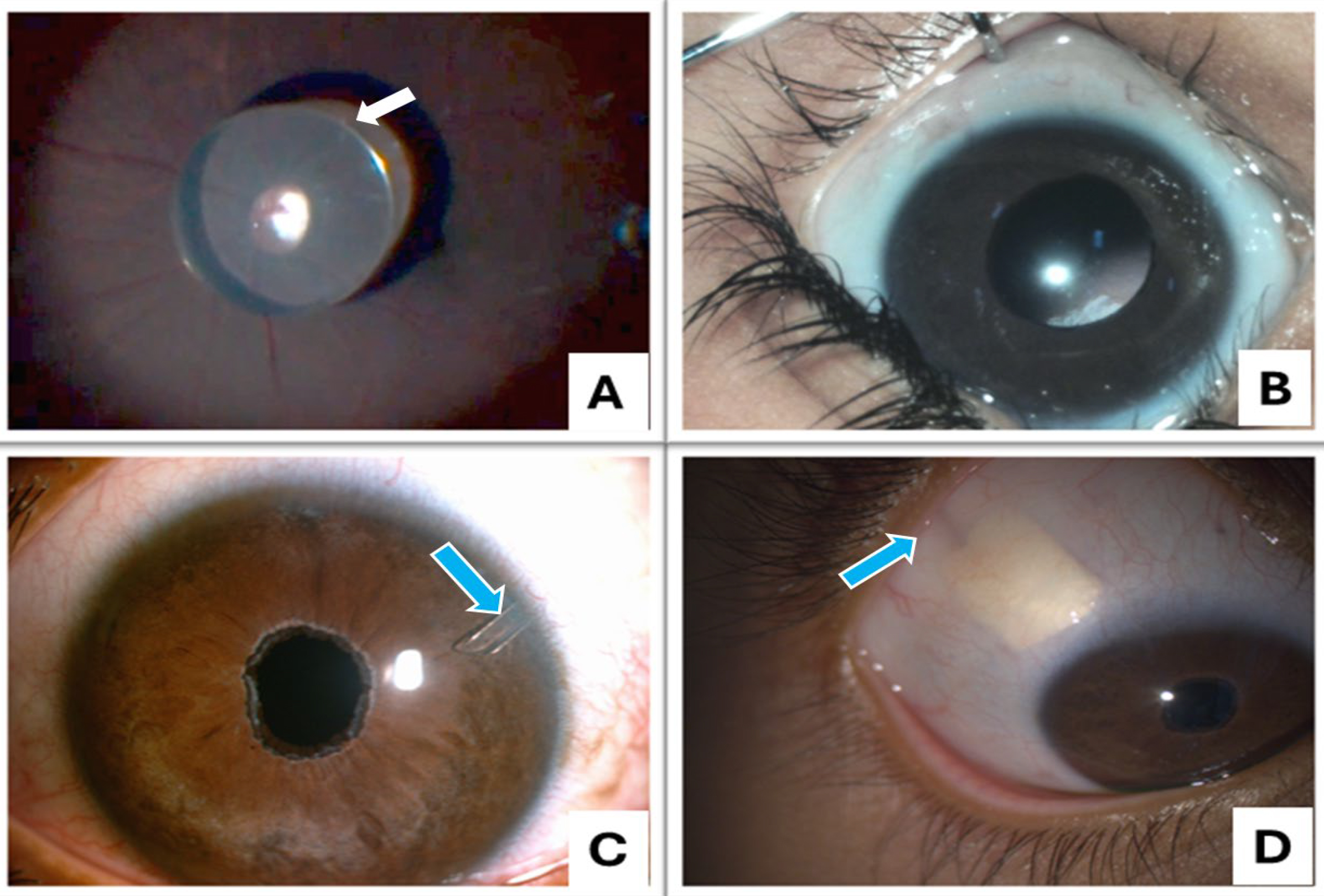
 Figure 3 of
Verma, Mol Vis 2025; 31:55-67.
Figure 3 of
Verma, Mol Vis 2025; 31:55-67.