Figure 2. The spectrum of ocular features observed in the LTBP2 patient’s cohort. A: Showing whitish pupillary border (possible persistent pupillary membrane (PPM) remnant, marked with arow), in right eye
of case P1. B: Indicating PPM in the the left eye of case P1. C: Showing Ectropion Uveae (EC) in case P13 (note a small area of whitish membrane in the area where EC has not developed yet
(marked with star). D: Showing anterior segment image with gross iridodonesis in an eye with posteriorly dislocated lens in case P17 (note the
shadow of the iridodonesis marked with arrow). E: Showing an anteriorly dislocated microspherophakic lens in the LE of case P12 wandering between anterior and posterior chamber,
(marked with star). F: Shows posteriorly dislocated lens in the RE of case P12, and G: Shows peripheral retinal lattice in case P2.
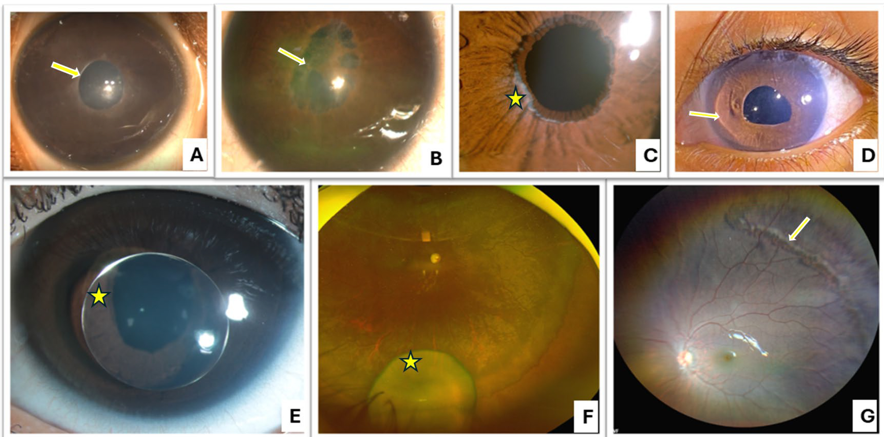
 Figure 2 of
Verma, Mol Vis 2025; 31:55-67.
Figure 2 of
Verma, Mol Vis 2025; 31:55-67.