Figure 5. SD-OCT imaging of patients carrying RP-associated gene variants: PRPF31 (P38), RPGR (P12), USH2A (P16), CRB1 (P24), RPGR (P11).
Panel A corresponds to the right eye (OD); panel B corresponds to the left eye (OS). Presentation of the ellipsoid zone (EZ):
In P38, the central macula showed a preserved EZ, whereas the perifoveal macula exhibited a not detected EZ in both eyes (OU);
in P12, the central macula displayed a disorganized EZ in OD and a not detected EZ in OS; the perifoveal macula showed a not
detected EZ in OU. Presentation of hyperreflective foci (HRF): In P16, HRF were observed in the perifoveal macula in OU; in
P24, HRF were observed in the central macula and in the perifoveal macula in OU. In P11, a lamellar macular hole was present
in the OD and an epiretinal membrane in the OS. RP, retinitis pigmentosa.
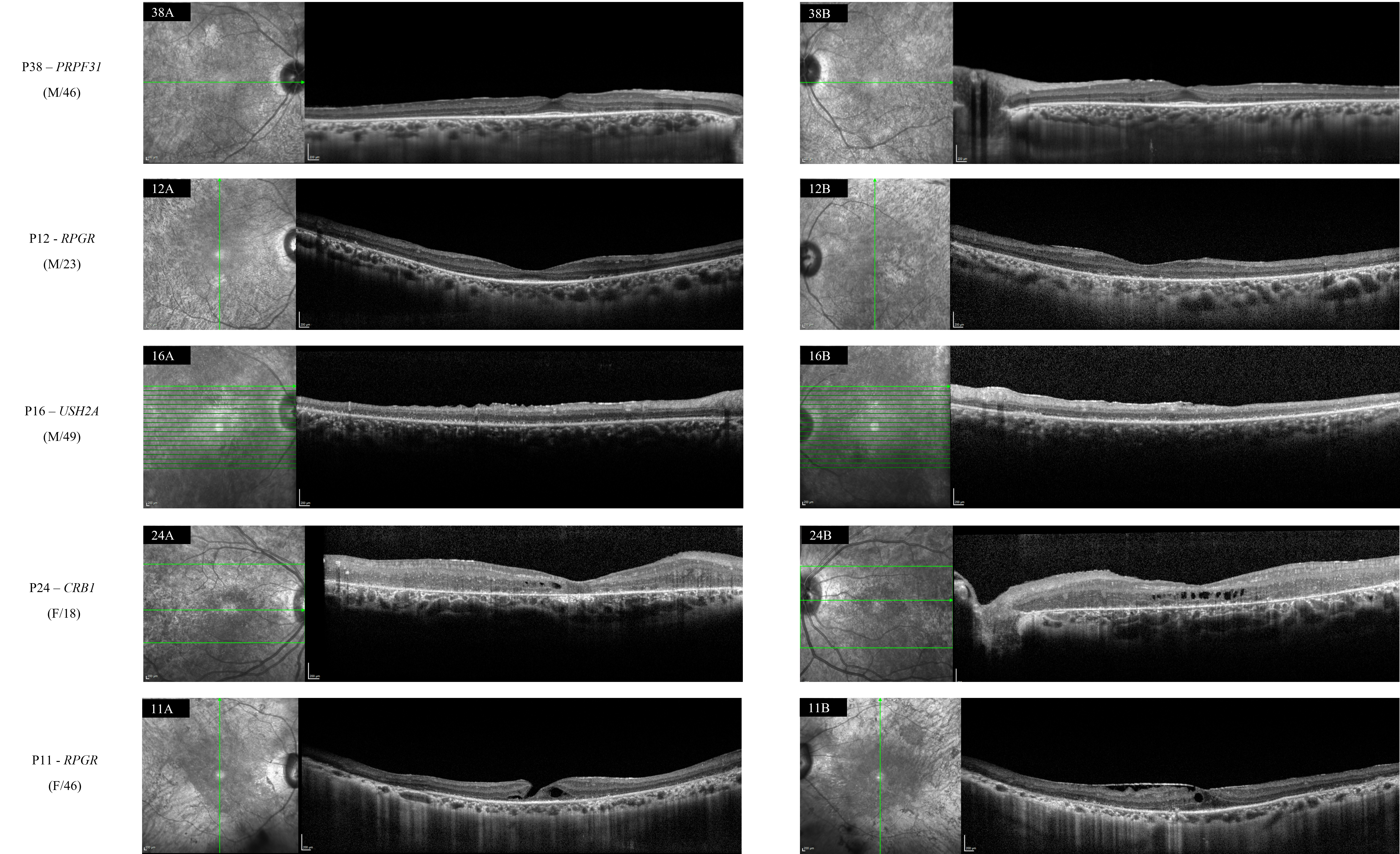
 Figure 5 of
de Freitas Cenachi, Mol Vis 2025; 31:526-537.
Figure 5 of
de Freitas Cenachi, Mol Vis 2025; 31:526-537.