Figure 4. Multimodal imaging of patients carrying RP-associated gene variants: PCARE (P31), PRPF8 (P37), EYS (P26, P28), and CHM. Images
include fundus autofluorescence (FAF; A and D) and fundus photography (B and C). Panels A and B correspond to the right eye;
panels C and D correspond to the left eye. In patients P31 (PCARE) and P37 (PRPF8), FAF images (P31A, P31D; P37A, P37D) show
hypoautofluorescent patches surrounding the arcades, corresponding to areas of retinal pigmentation and atrophy that may be
less evident on color fundus photography (P31B, P31C; P37B, P37D). In patients P26 and P28, both harboring EYS variants, distinct
FAF patterns previously associated with this gene were observed: a “broad-banded hyperautofluorescent leading edge” in P26
and an “infinity sign” in P28. Fundus photographs of these patients demonstrate peripheral pigmentation, vascular attenuation,
and optic disc pallor in both patients. In the patient with a pathogenic CHM variant (choroideremia), FAF imaging revealed
scalloped mid-peripheral chorioretinal atrophy—a hallmark feature of the disease. Fundus photography displayed overlapping
features with RP, including peripheral pigmentation, vessel narrowing, and optic disc pallor. RP, retinitis pigmentosa.
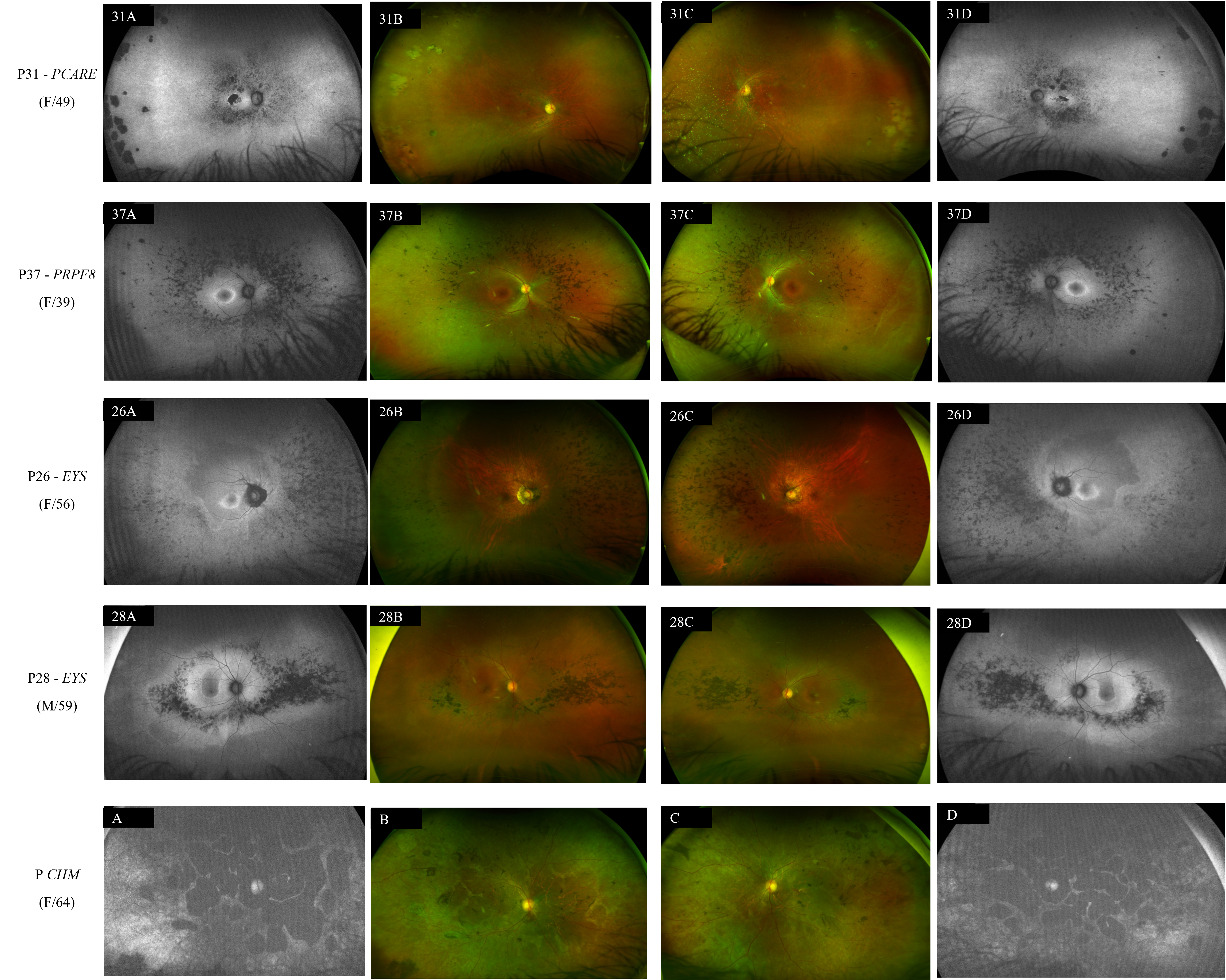
 Figure 4 of
de Freitas Cenachi, Mol Vis 2025; 31:526-537.
Figure 4 of
de Freitas Cenachi, Mol Vis 2025; 31:526-537.