Figure 3. Genetic and clinical characterization of patients with the RHO gene variant in the cohort. The patients harbor three different
variants: c.601G>T in P1, c.316G>A in P2, and c.403C>T in P4 to P8, the latter being related patients (P4 is the sibling of
P5, who is the mother of P6, P7, and P8). Images include color fundus photography (A), fundus autofluorescence (FAF; B), and
optical coherence tomography (OCT; C) of the right eye. Color fundus photographs show peripheral pigmentation, vascular attenuation,
and optic disc pallor in both eyes of all patients. FAF imaging highlights the presence of the parafoveal hyperautofluorescent
ring (e.g., P1B, P6B, P7B, P8B), corresponding on OCT to the demarcation between areas of central photoreceptor preservation
and regions where the outer retina is structurally disrupted (e.g., P1C, P6C, P7C, P8C). Additional OCT findings include large
cysts in the internal and external layers, and surrounding hyperreflective plaques in the outer retina in P5C correspond to
a macroaneurysm. In 8C, cysts are identified in both the inner and outer retinal layers. This figure illustrates phenotypic
heterogeneity: patient P2, who carries the c.316G>A variant, exhibited sectoral retinal involvement, whereas other patients
with different variants in the same gene showed diffuse retinal involvement. Clinical heterogeneity is also evident, as patient
P8, aged 9 years, carries the same mutation as her older sisters but presents with more pronounced macular edema.
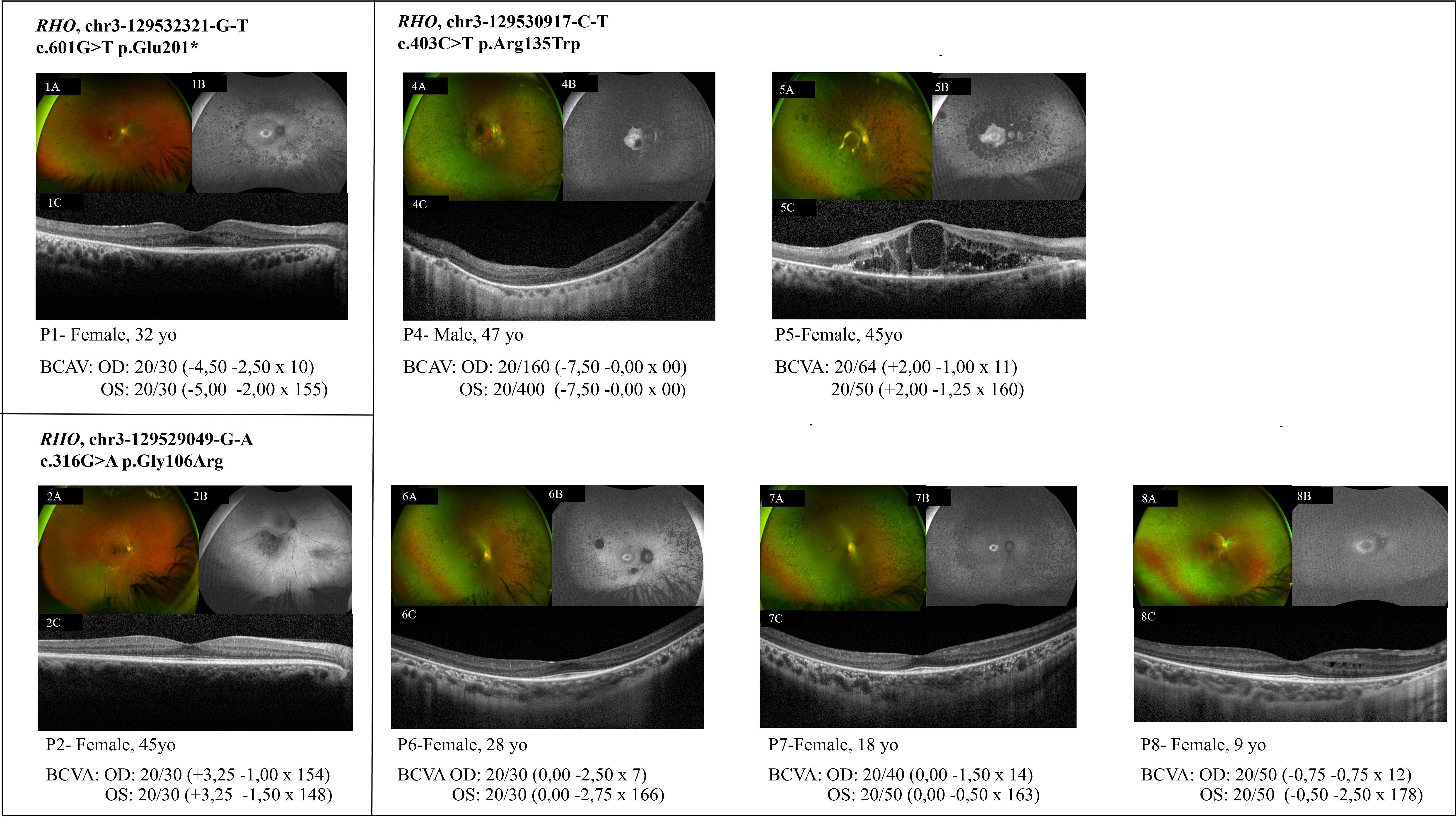
 Figure 3 of
de Freitas Cenachi, Mol Vis 2025; 31:526-537.
Figure 3 of
de Freitas Cenachi, Mol Vis 2025; 31:526-537.