Figure 4. Fundus autofluorescence (FAF) and optical coherence tomography (OCT) of both eyes for P29 and P7. A. Images of P29 demonstrate peripheral hypoautofluorescence extending beyond the vascular arcades and a hyperautofluorescent
ring at the transition zone on FAF. OCT images reveal foveal schisis (white arrows), consistent with concentric peripheral
retinal degeneration characteristic of enhanced S-cone syndrome. B. P7 shows bilateral retinitis pigmentosa with widespread FAF abnormalities and cystoid macular edema on OCT (red arrows),
indicating intraretinal fluid accumulation and advanced retinal dysfunction.
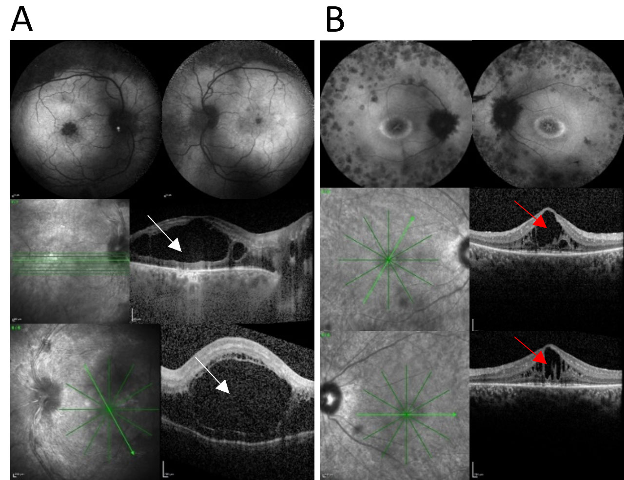
 Figure 4 of
Keles, Mol Vis 2025; 31:502-513.
Figure 4 of
Keles, Mol Vis 2025; 31:502-513.