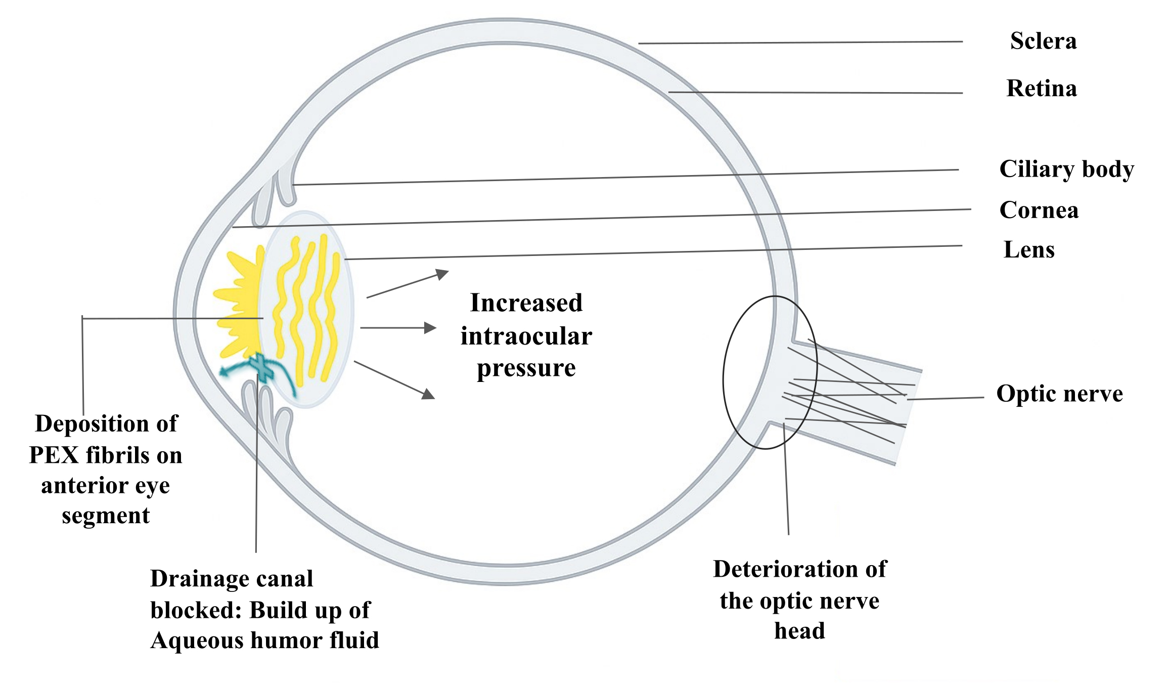
Figure 1. Schematic representation of pseudoexfoliation pathology in the human eye. Yellow fibrillar deposits on the anterior lens capsule
and pupillary margin represent PEX material, which can obstruct aqueous humor outflow, leading to increased intraocular pressure
and subsequent glaucomatous optic nerve damage. The diagram illustrates key ocular structures involved in the disease process.
Outline of the eye adapted from
OpenClipart.
 Figure 1 of
Hayat, Mol Vis 2025; 31:463-484.
Figure 1 of
Hayat, Mol Vis 2025; 31:463-484.