Figure 2. Examples of multimodality images in three patients with achromatopsia. Patient 1 demonstrates central hyperautofluorescence of
the fovea. A: Infrared (IR) shows a normal reflectivity pattern. B: OCT shows stippling of the ellipsoid zone. C: No outer segment loss or other abnormalities appear in the retinal layers. Patient 2 demonstrates a normal autofluorescence
pattern. D: IR shows central foveal hyper-reflectance. E: OCT shows stippling of the ellipsoid zone. F: No outer segment loss or other abnormalities appear in the retinal layers.
Patient 3 shows central foveal hypoautofluorescence with a hyperautofluorescent halo. G: IR shows hyper-reflectance at the fovea lesion. H: OCT demonstrates foveal outer retina atrophy with secondary choroidal hyperreflectivity. I: Retinal layers outside the fovea are well preserved.
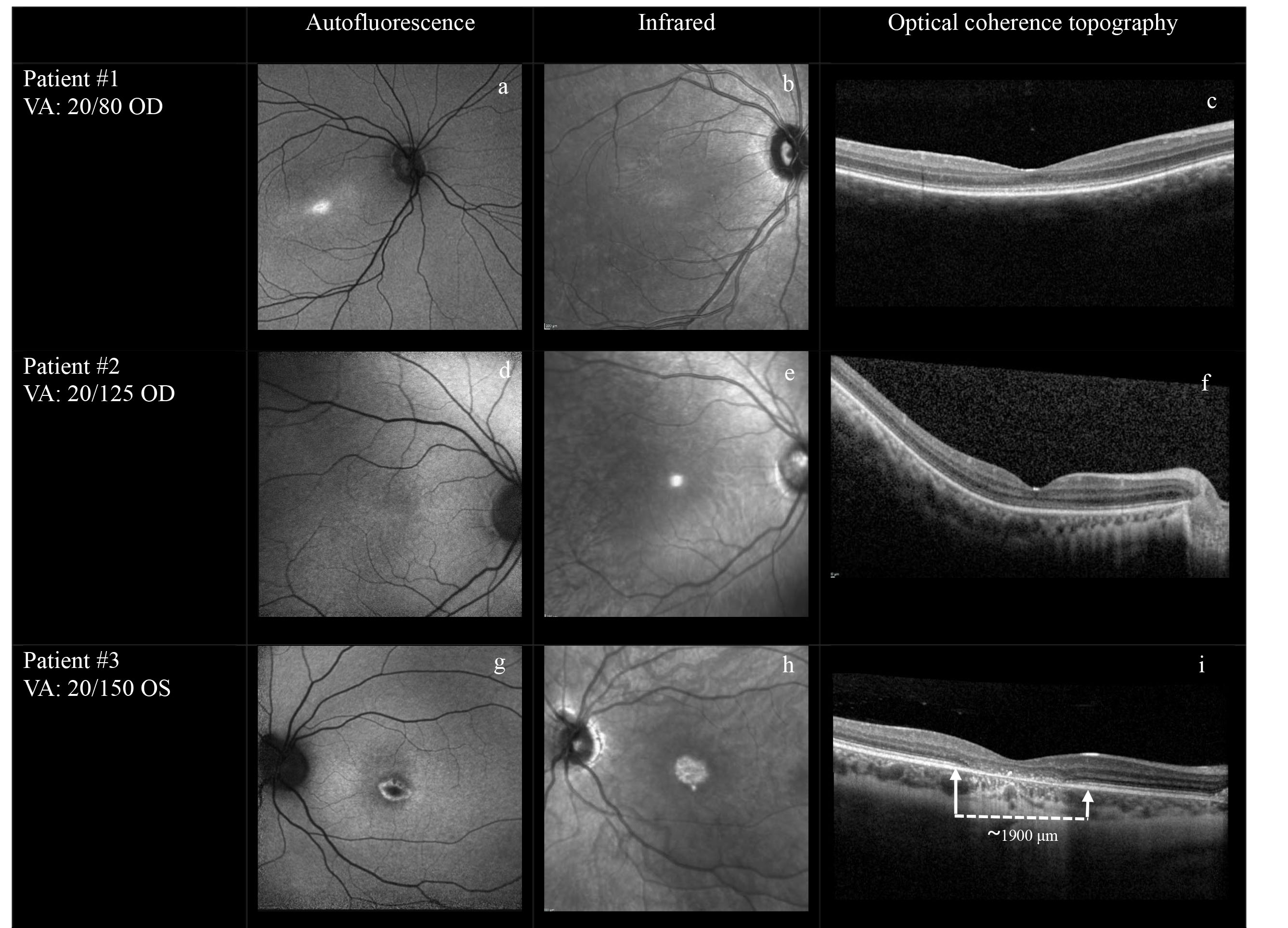
 Figure 2 of
Molleti, Mol Vis 2025; 31:454-461.
Figure 2 of
Molleti, Mol Vis 2025; 31:454-461.