Figure 5. Images and full-field electroretinogram of Case 6 with RPE65 gene mutation. Case 6 shows a proband with mutation c.11+5 G>A and c.1520C>T; p.(Ala507Val) at the age of 12 years. The fundus
(A and D) shows diffuse pigmentary changes in the retina, and blood vessels, optic disc, and macula apparently preserved in both eyes
(OU). OU optical coherence tomography (B and E) shows subfoveal outer retina preserved, and perifoveal atrophy with loss of the ellipsoid zones. In the left eye (OS) vitreous
cavity, there are signs of vitreous degeneration (highlighted by the orange arrow). The full-field electroretinogram shows
minimal rod and cone responses in OU (C and F).
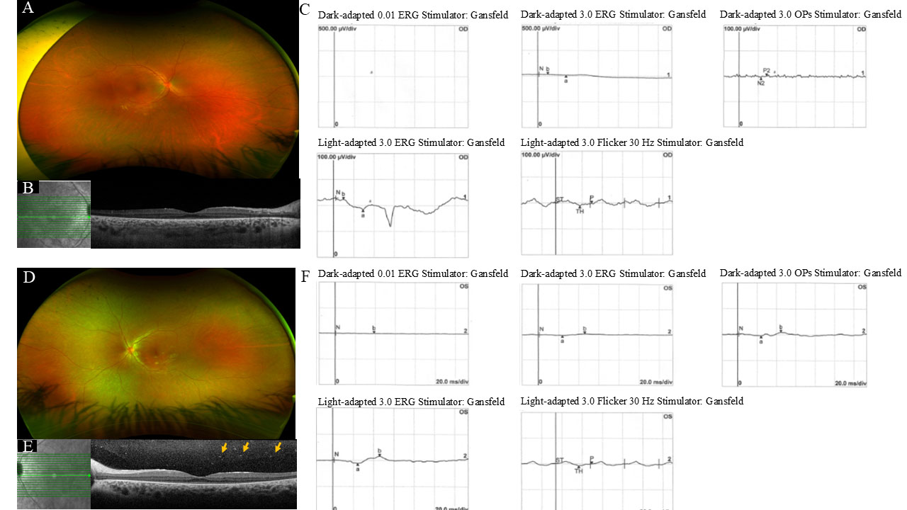
 Figure 5 of
de Freitas Cenachi, Mol Vis 2025; 31:45-54.
Figure 5 of
de Freitas Cenachi, Mol Vis 2025; 31:45-54.