Figure 3. Images and full-field electroretinogram of Case 4 with RPE65 gene mutation. Case 4 shows a proband with mutation c.1022 T>C;p.(Leu341Ser) and c.560G>A;p.(Gly187Glu) in the RPE65 gene at the age of 20 years with fundus (A and D) showing severe central macular atrophy (highlighted by the white arrows), slight pallor of the optic disc, attenuation of
blood vessels, and diffuse retinal pigment epithelium mottling degeneration. Optical coherence tomography shows severe overall
retinal thinning and photoreceptor loss in the macula (B and E). The full-field electroretinogram shows minimal rod and cone responses in right and left eyes (C and F).
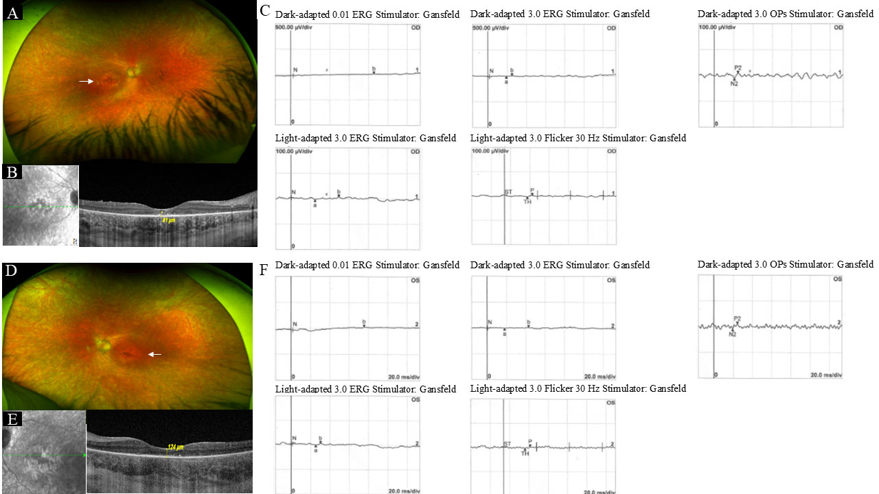
 Figure 3 of
de Freitas Cenachi, Mol Vis 2025; 31:45-54.
Figure 3 of
de Freitas Cenachi, Mol Vis 2025; 31:45-54.