Figure 2. Images of Cases 1, 2 and 3 with RPE65 gene mutation. Cases 1, 2, and 3 the variant c.1022T>C; p.(Leu341Ser) was identified in the RPE65 gene. Case 1. Fundus of the 25-year-old sibling shows (A and D) attenuation of blood vessels, diffuse mottling of the retinal pigment epithelial with subtle pigments, slight pallor of
the optic disc, and macular atrophy (highlighted by the white arrows). The fundus autofluorescence of both eyes (OU) shows
diffuse background hypo-autofluorescence (B and E). Optical coherence tomography (OCT) of the OU (C and F) shows thinning of the outer retina and total loss of the ellipsoid zone of photoreceptors. Case 2. The proband at the age
of eight years, with the fundus (G and J) showing subtle granular retinal pigment epithelium degeneration. The optic discs, the blood vessels, and the macula appear
to have no clinically significant change. Fundus autofluorescence of OU presents an insufficient image for analysis of possible
retinal changes (H and K). The OCT (I and L) shows sparse disruptions in the ellipsoidal zone and outer nuclear membrane (highlighted by the orange arrows). Case 3.
The three-year-old sibling with the right eye (OD) fundus presenting pigmentary mottling in the mid-peripheral retina (M, highlighted by the yellow arrow) and optic disc, blood vessels, and macula with no notable change. The fundus autofluorescence
shows an insufficient image for analysis (N). The OCT (O) shows no changes in retinal thickness.
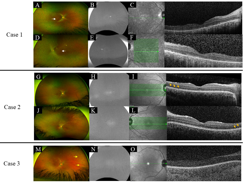
 Figure 2 of
de Freitas Cenachi, Mol Vis 2025; 31:45-54.
Figure 2 of
de Freitas Cenachi, Mol Vis 2025; 31:45-54.