Figure 4. Functional food mix preserved retinal architecture and loss of retinal cells in streptozotocin induced diabetes. A: Hematoxylin and eosin staining of rat retinal sections. B: Immunofluorescence staining of rhodopsin in the retina. Rhodopsin stain was reduced in the D group and improved with FF
supplementation. Blue: Nuclear stain with 4′,6-diamidino-2-phenylindole. Red: Rhodopsin stain. Merged: 4′,6-diamidino-2-phenylindole
and rhodopsin. 40× magnification, scale bar = 10 µm, and n = 3 per group. C, control; D, diabetes (untreated); D+FF1, diabetes
treated with FF1; D+FF2, diabetes treated with FF2 group; GCL, ganglion cell layer; INL, inner nuclear layer; IPL, inner plexiform
layer; ONL, outer nuclear layer; OPL, outer plexiform layer; PRL, photoreceptor layer; RPE, retinal pigment epithelium layer.
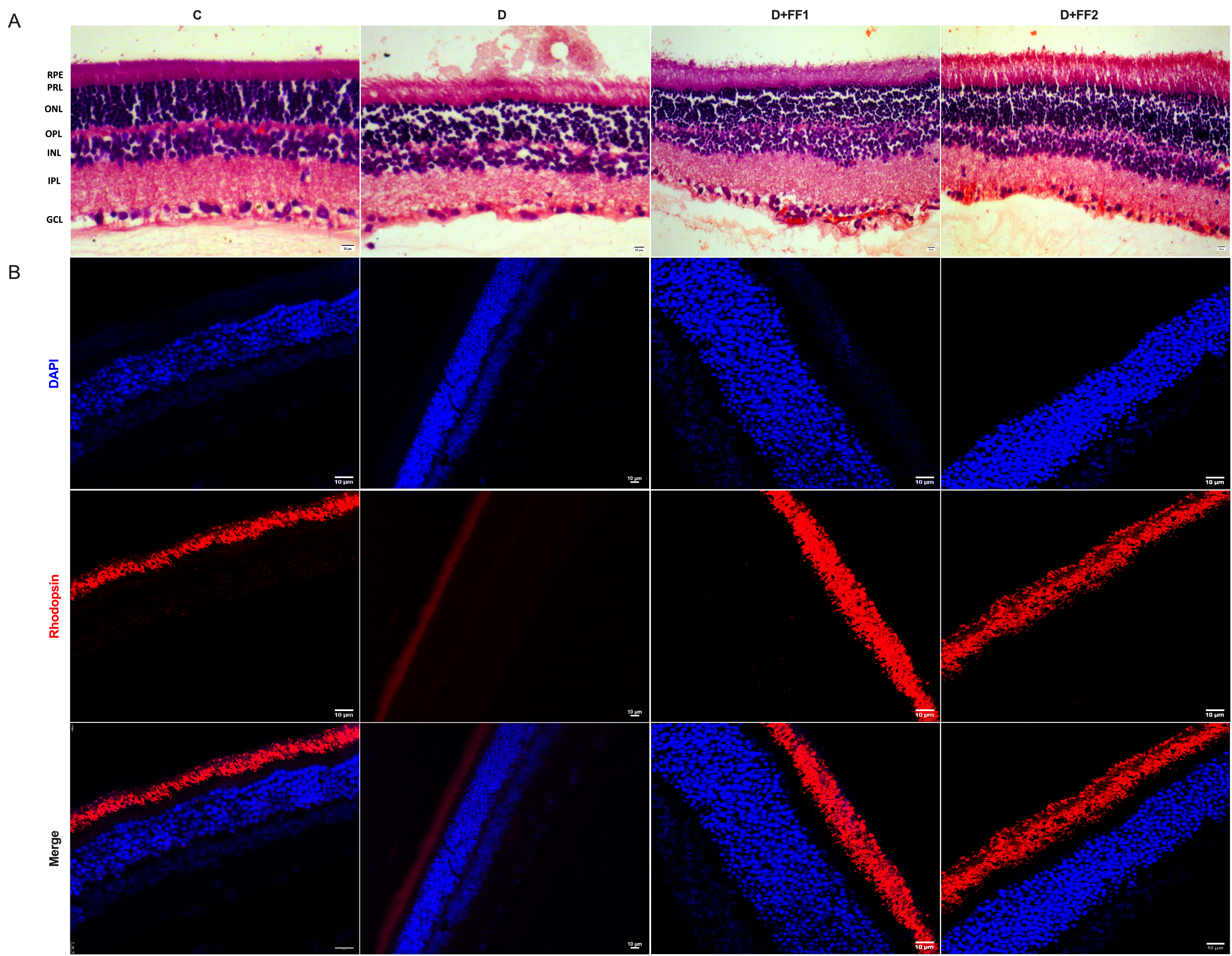
 Figure 4 of
Kalahasti, Mol Vis 2025; 31:411-422.
Figure 4 of
Kalahasti, Mol Vis 2025; 31:411-422.