Figure 2. Vitreous samples obtained from patients with diabetic macular edema (DME) and proliferative diabetic retinopathy (PDR) modulate
the activation markers of peripheral blood neutrophils. The upper panels show histograms of the expression of CD11b (A), CD15 (B), and CD66b (C) on neutrophils incubated with the vitreous of surrogate control (blue), DME (green), or PDR (red) patients. The lower panels
are bar graphs of the medium fluorescence intensity (MFI) of the activation markers CD11b (A), CD15 (B), and CD66b (C) in peripheral neutrophils incubated with the vitreous of surrogate control (white bars), DME (gray bars), or PDR (black
bars) patients. We found a significant decrease in both CD11b and CD15 activation markers in neutrophils exposed to the vitreous
from patients with PDR. The bars represent the means ± standard error of the means. *p < 0.05. **p < 0.01.
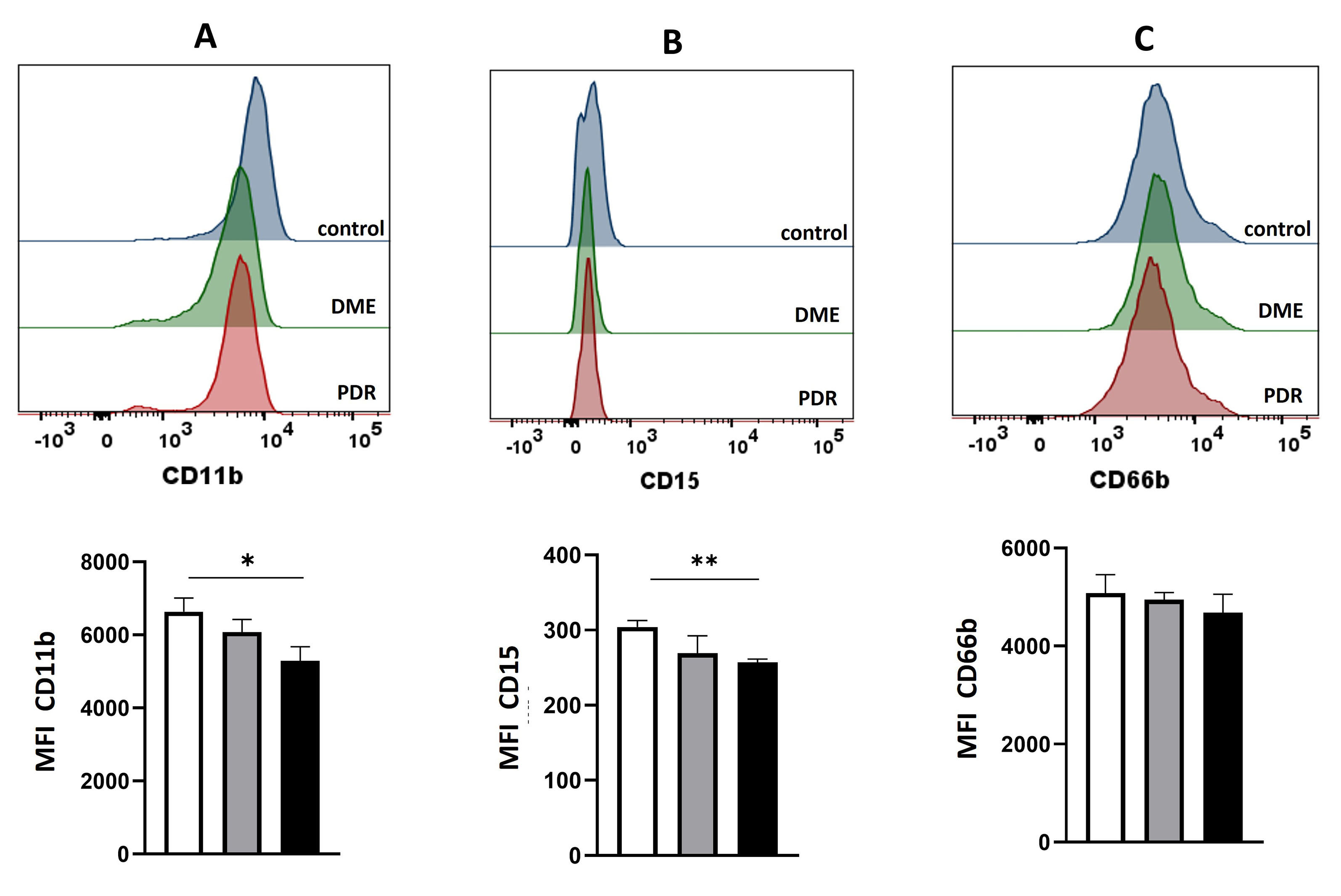
 Figure 2 of
Magaña-Guerrero, Mol Vis 2025; 31:368-378.
Figure 2 of
Magaña-Guerrero, Mol Vis 2025; 31:368-378.