Figure 6. Characteristics of foveal development in the anole retina. Comparison of the embryonic stage (ES) panels ES7 and ES10 (top
row) show that at ES10, the ganglion cell layer (GCL) is thickened (green arrow) compared to the retina at embryonic stage
ES7. INL, inner nuclear layer (size bar = 50 μm). The start of foveal pit formation is shown in the ES17 panel (arrow), where
the fovea has a thickened outer nuclear layer (ONL), compared to a one cell thick ONL at ES10. Comparing the adult and human
panels (bottom row), the adult anole fovea has a thick nerve fiber layer (NFL), and there are no nuclei in the ONL in the
center of the fovea (size bar = 100 μm) compared to the human foveola, where cells in the ONL are present. When the retina
is removed from the posterior anole eye cup, a yellow pigment is revealed (bottom row, third image). The red dotted line shows
where the foveal pit (FP) would be positioned, and the yellow pigment extends beyond the FP (white dotted line). The immunochemistry
images on the top and bottom rows show that Pax6 protein expression (green) is highest at ES17 in the GCL and at lower levels
in the INL compared to ES14 (size bar = 100 μm). The retinal sections are counterstained with Hoechst dye to identify the
three nuclear layers. The far-right panel is a Western blot with quantification of signal intensity plot below, showing a
nearly three-fold increase in pigment epithelial-derived factor (PEDF) in foveal (F) retina at ES10 compared to non-foveal
(NF) retina (p < 0.001, n = 4), which is not significantly elevated at ES17. Loading control is GAPDH.
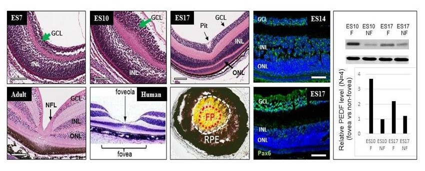
 Figure 6 of
Gregory-Evans, Mol Vis 2025; 31:319-343.
Figure 6 of
Gregory-Evans, Mol Vis 2025; 31:319-343.