Figure 3. Imaging of a female ocular albinism carrier with confirmed genetic testing (GPR143 variant). Fundus images are captured using
Optos ultra-widefield color imaging in the patient’s left (A) and right (B) eyes. Fundus autofluorescence imaging in the patient’s left (C) and right (D) eyes, showing abnormal pigmentary streaks in the peripheral retina. Optical coherence tomography imaging in the patient’s
left (E) and right (F) eyes, showing partial foveal hypoplasia. A shallow pit is seen, with the absence of the expansion of the outer nuclear layer
seen in normal fovea. The green line indicates where the foveal thickness was measured (the left eye was 185 μm thick and
the right eye was 182 μm thick. The normal thickness of the fovea is ≥210 μm).
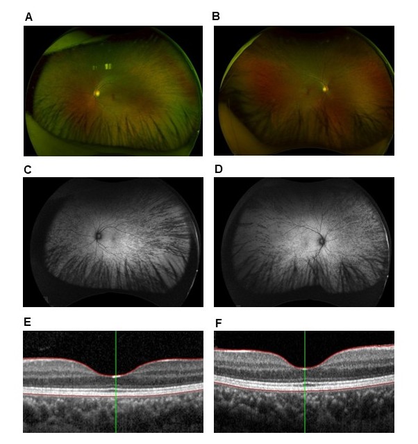
 Figure 3 of
Gregory-Evans, Mol Vis 2025; 31:319-343.
Figure 3 of
Gregory-Evans, Mol Vis 2025; 31:319-343.