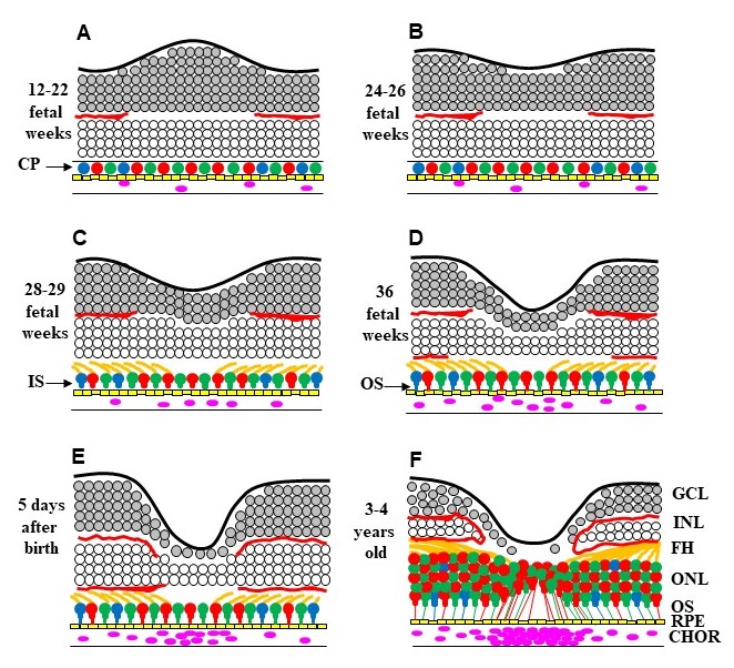
Figure 2. A representative schematic of the different stages of foveal development.
A: This image shows ganglion cell layer (GCL) thickening at the position of the future foveal pit and ingrowth of blood vessels
(red lines) between 12 and 22 weeks. Only a single row of cone photoreceptor (CP) cells is present.
B: In this image the foveal depression starts to form between 24 and 26 weeks.
C: In this image the GCL and inner nuclear layer (INL) start to thin out, and the fibers of Henle (FH) and the inner segments
(ISs) of CP are formed.
D: This image shows further excavation of the GCL and INL resulting in a deeper foveal pit, and the outer segments (OSs) of
the CP are starting to form.
E: This image shows the fovea shortly after birth, where the GCL and INL are one-to-two cells thick. Throughout development,
the blood vessels do not encroach upon the foveal retina, forming the foveal avascular zone.
F: This image shows the fovea by the fourth year of life, where the foveal region is packed with CPs in the outer nuclear layer
(ONL) with elongated OSs. The choroid (CHOR) is filled with blood vessels to supply the outer retina and retinal pigment epithelium
(RPE) with oxygen and nourishment. This is adapted from a previous publication by the authors [
1].
 Figure 2 of
Gregory-Evans, Mol Vis 2025; 31:319-343.
Figure 2 of
Gregory-Evans, Mol Vis 2025; 31:319-343.