Figure 4. The figure shows the ophthalmologic findings of the index case in family 3. Optic disc photographs of the right eye (RE) and
left eye (LE) show a relatively preserved neuroretinal rim in both eyes (A). Retinal nerve fiber layer (RNFL) analysis performed with Cirrus optical coherence tomography (OCT) reveals a superior defect
in the RE, indicating early RNFL loss consistent with mild to moderate glaucoma in this eye (B). LE, left eye; RE, right eye; RNFL, retinal nerve fiber layer.
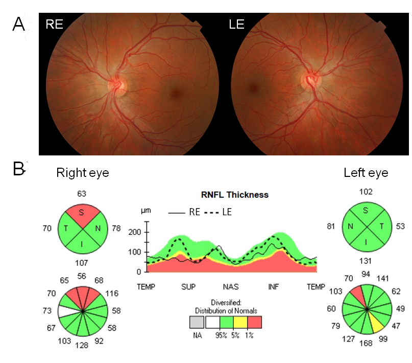
 Figure 4 of
Laguna, Mol Vis 2025; 31:306-317.
Figure 4 of
Laguna, Mol Vis 2025; 31:306-317.