Figure 3. Fundoscopic examination of the index case in family 2 showed optic discs with well-defined margins that were somewhat pale,
with optic disc cupping of 0.5 in the right eye (RE) and 0.7 in the left eye (LE). The superior neuroretinal rim was decreased
in the RE and diffusely thinned in the LE. No hemorrhages were observed. These findings are consistent with moderate-to-severe
glaucoma. Drusen were dispersed in the posterior pole. The RE displayed filiform vessels and had undergone panretinal photocoagulation
due to a previous central retinal vein occlusion. The RE presents filiform vessels and underwent panretinal photocoagulation
due to a previous central retinal vein occlusion (RE). LE, left eye; RE, right eye.
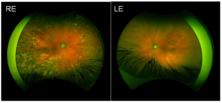
 Figure 3 of
Laguna, Mol Vis 2025; 31:306-317.
Figure 3 of
Laguna, Mol Vis 2025; 31:306-317.