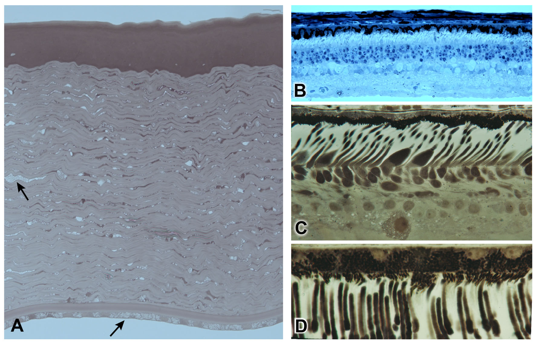
Figure 1. Structural appearance of the archival tissue sections from which the plastic-embedded tissue blocks were used for DNA extraction.
Sections were stained with paraphenylenediamine (PPD;
A,
C,
D) or azure II/methylene blue (
B).
A: A corneal section from dog CLF4 affected with mucopolysaccharidosis VII shows the typical vacuolated inclusions in the stromal
keratocytes and corneal endothelial cells [
16].
B: A mid-peripheral retinal section from
prcd-affected dog CLF1 shows disorganization of the photoreceptor layer and ~50% reduction in outer nuclear layer thickness [
17].
C,
D: Panels show images of the far peripheral (
C) retina of normal nonhuman primate MR1 and the photoreceptor outer segment–retinal pigment epithelial interface of the mid-peripheral
retina (D).
 Figure 1 of
Niggel, Mol Vis 2025; 31:297-304.
Figure 1 of
Niggel, Mol Vis 2025; 31:297-304.