Figure 4. Color fundus and autofluorescence images from affected participant III-1. The upper row shows color fundus images from age
63 years showing bone spicule retinal degeneration confined to the near periphery. The bottom row shows autofluorescence imaging
showing a hypo-autofluorescence band confined to the posterior pole plus hyper-autofluorescence beyond this area and at each
macula. This would be described as pericentral retinitis pigmentosa.
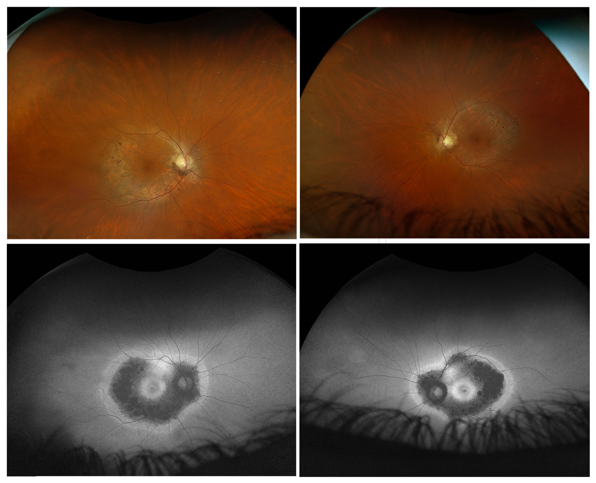
 Figure 4 of
Gregory-Evans, Mol Vis 2025; 31:175-188.
Figure 4 of
Gregory-Evans, Mol Vis 2025; 31:175-188.