Figure 3. Deidentified Goldmann visual field results from affected participants III-1 and III-4. The upper row shows results from participant
III-1 at 61 years of age showing dense mid-peripheral ring scotomas extending from 5 to 30 degrees. The middle row shows results
from participant III-4 at 43 years old showing extensive field loss beyond the mid-periphery. The bottom row shows results
from participant III-4 at 55 years of age showing further decline of far peripheral islands of vision.
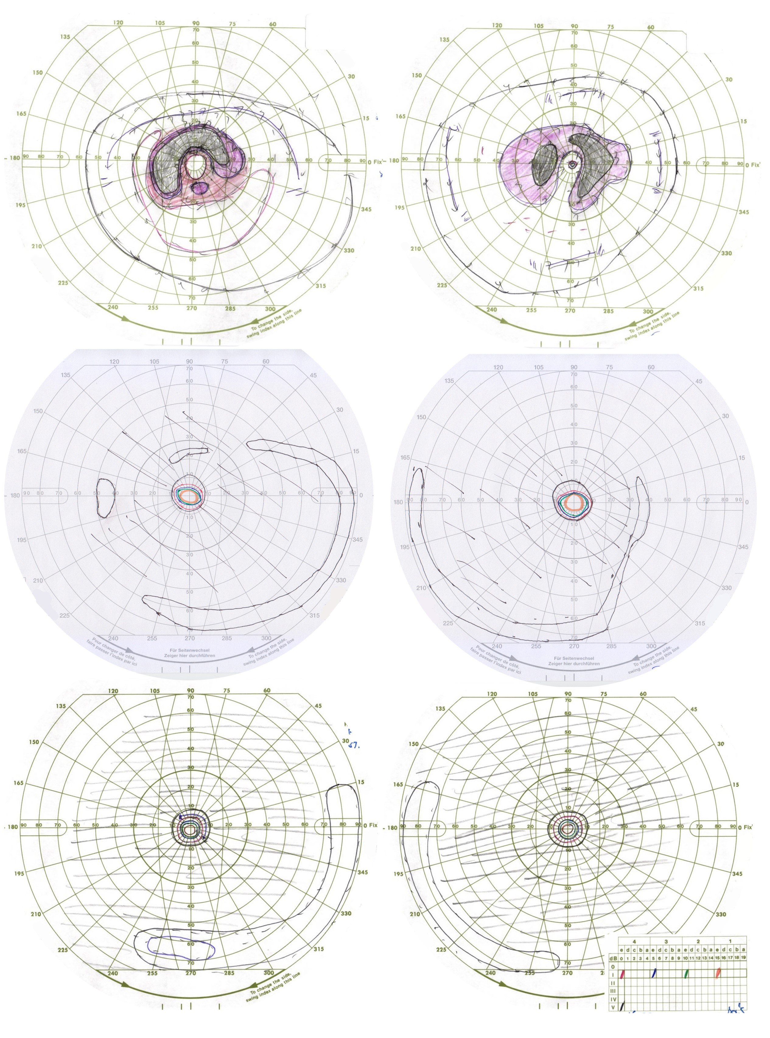
 Figure 3 of
Gregory-Evans, Mol Vis 2025; 31:175-188.
Figure 3 of
Gregory-Evans, Mol Vis 2025; 31:175-188.