Figure 2. Color fundus and autofluorescence images from affected participant III-4. The upper row are color fundus images from age 51
years showing bone spicule retinal degeneration extending into the peripheral retina. The middle row shows color fundus images
from age 62 years showing further extension of bone spicule retinal degeneration into the macular area and peripheral retina.
Bottom row, autofluorescence imaging showing widespread hypo-autofluorescence plus hyper-autofluorescence at each macula.
This would be described as typical retinitis pigmentosa.
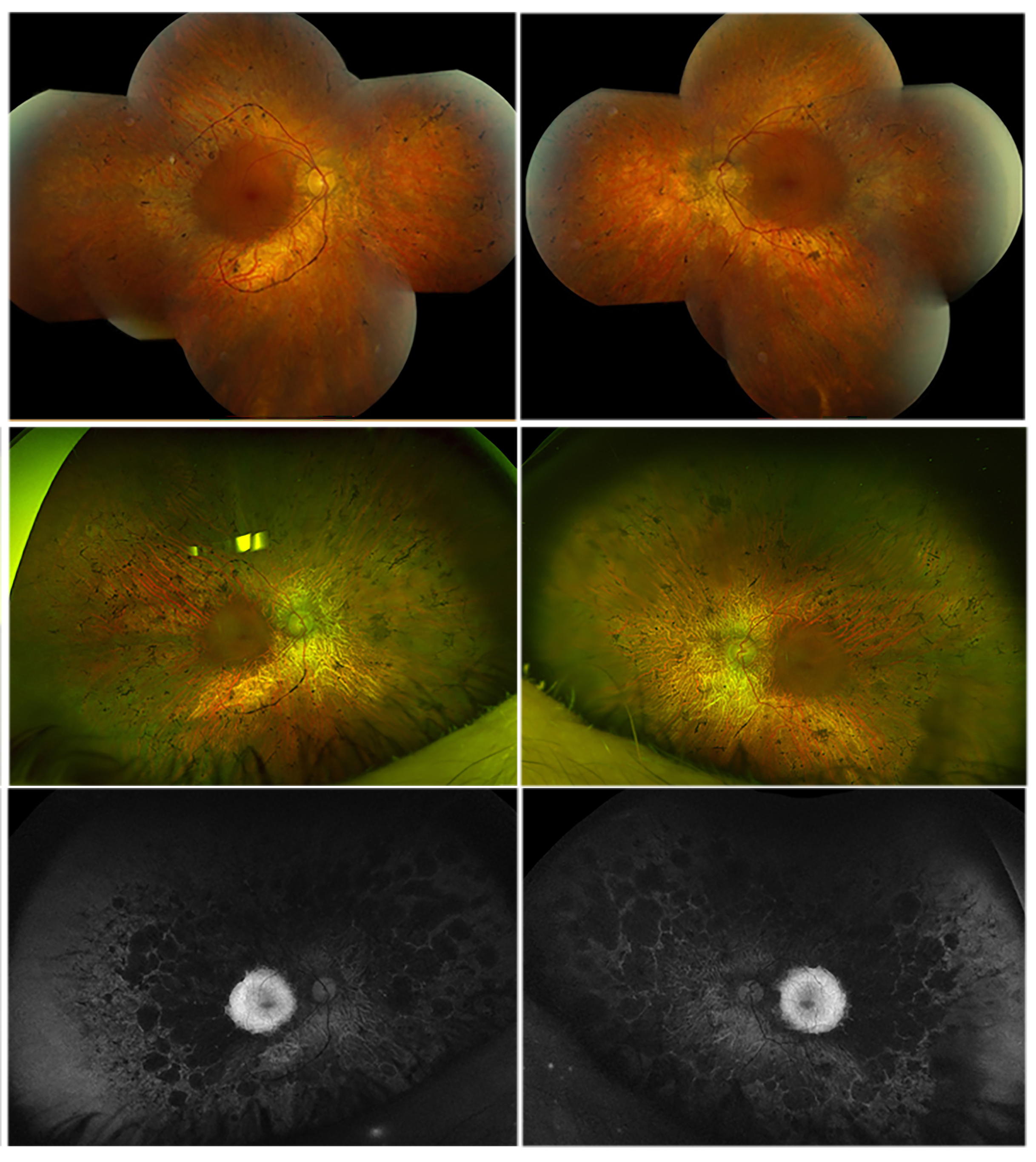
 Figure 2 of
Gregory-Evans, Mol Vis 2025; 31:175-188.
Figure 2 of
Gregory-Evans, Mol Vis 2025; 31:175-188.