Figure 1. Retinal neovascularization and formation of nonperfusion areas in each group. Compared with the normal group, the retinal
veins in the oxygen-induced retinopathy group are beaded, with a large avascular perfusion area in the center (triangle) and
a large number of highly fluorescent stained neovascularization groups are visible in the surrounding area (arrow).
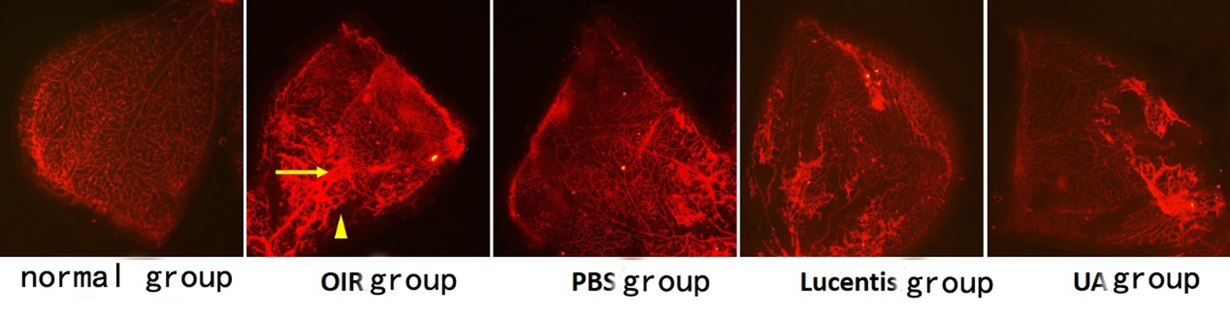
 Figure 1 of
Yang, Mol Vis 2025; 31:160-173.
Figure 1 of
Yang, Mol Vis 2025; 31:160-173.