Figure 9. Fundus images of the rabbits. A: Total retinal detachment; the optic disc is indicated by an arrow (↑;subject no. C1, stage 5). B: Focal traction and intravitreal membrane formations and gliotic changes in the retina (subject no. C2, stage 2). C: Traction creating a membrane (↑) and a wide detachment area (De; subject no. C4, stage 4). D: Intravitreal membrane, vitreous opacities, and gliotic changes in the retina (subject no. N1, stage 1). E: Membrane extending from the disc (OD) and that does not produce traction, indicated by an arrow (↑;subject no. N2, stage
1). (F) Intravitreal membrane indicated by an arrow (↑), and gliotic changes in the retina (subject no. N6, stage 1).
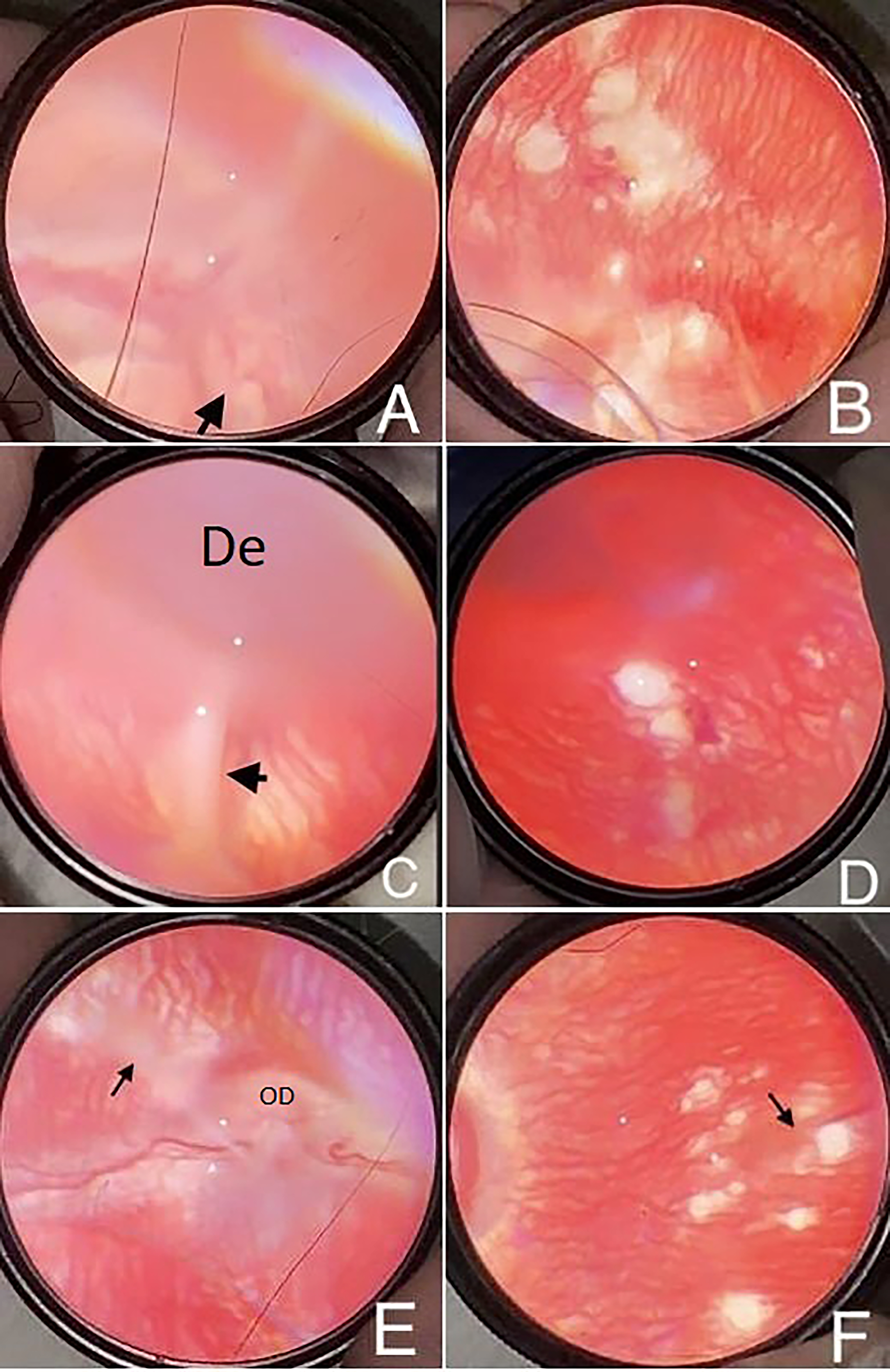
 Figure 9 of
Arslan, Mol Vis 2025; 31:142-157.
Figure 9 of
Arslan, Mol Vis 2025; 31:142-157.