Figure 4. Restoration of PDE6B expression and preservation of photoreceptor cells in Pde6b knockout rats following AAV5-Pde6b treatment. A: Experimental scheme. B: Reverse transcriptase PCR analysis of the retinal homogenates from wild-type (WT) and Pde6b knockout (KO) rats injected with AAV5-GFP or AAV5-Pde6b (n=4 for each group). C–E: Representative images of PDE6B and Rhodopsin in the retina from WT (C, F) and Pde6b KO rats injected with AAV5-GFP (D, G) or AAV5-Pde6b (E, H; n=4 for each group). F–H: Boxed areas on the merged panels are shown at a higher magnification on the left panel. I. Quantification of PDE6B staining
intensity in the photoreceptor outer segment. J: Quantification of Rhodopsin staining intensity in the photoreceptor outer segment. 4’,6-diamidino-2-phenylindole nuclear
counterstain is shown in blue. a.u., arbitrary units; GCL, ganglion cell layer; INL, inner nuclear layer; ONL, outer nuclear
layer; IS/OS, inner and outer segments; RPE, retinal pigment epithelium. Scale bar, 50 μM. Error bars, mean ± SEM, **p<0.01,
ANOVA with Student–Newman–Keuls post hoc analysis.
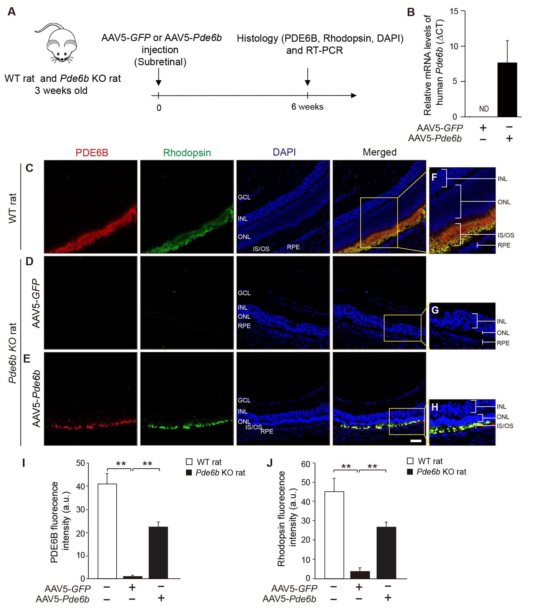
 Figure 4 of
Kim, Mol Vis 2025; 31:127-141.
Figure 4 of
Kim, Mol Vis 2025; 31:127-141.