Figure 3. AAV5-Pde6b rescues clinical phenotype in Pde6b gene knockout rats. A: Schematic diagram of the AAV-Pde6b construct. B: Experimental scheme. C–E: Representative color fundus photographs of wild-type rats (C) and Pde6b knockout rats injected with AAV5-GFP (D) or AAV5-Pde6b (E; n=4 for each group). F–H: Total retinal artery and vein analysis in the same rat retina as C, D and E, respectively. Retinal vessel diameters were measured by identifying the vessels marked in red within a yellow circle located
at a distance of 1 mm from the optic nerve head in the retina. I: Average retinal vessel diameters. Error bars, mean ± SEM, *p<0.05, ANOVA with Student–Newman–Keuls post hoc analysis.
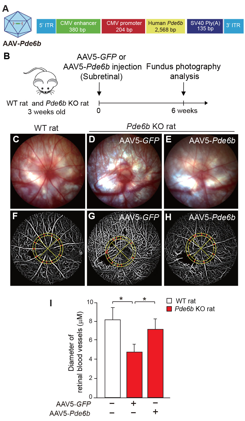
 Figure 3 of
Kim, Mol Vis 2025; 31:127-141.
Figure 3 of
Kim, Mol Vis 2025; 31:127-141.