Figure 2. Comparison of green fluorescence protein expression in wild-type and Pde6b knockout rat retinas after intravitreal and subretinal injection of different adeno-associated virus vectors. A, B: Green fluorescence protein (GFP) expression in the retinas of wild-type (WT; A) and Pde6b knockout (KO) rats (B) after intravitreal and subretinal injection of AAV2-GFP, AAV5-GFP and AAV8-GFP vectors at six weeks (n=4 for each group). 4’,6-diamidino-2-phenylindole nuclear counterstain is shown in blue. C, D: Quantitative analysis of AAV2-GFP, AAV5-GFP and AAV8-GFP fluorescence intensity on histological sections in the eyes of WT (C) and Pde6b KO rats (D). a.u., arbitrary units; GCL, ganglion cell layer; INL, inner nuclear layer; ONL, outer nuclear layer; IS, inner
segments; OS, outer segments; RPE, retinal pigment epithelium. Scale bar, 50 μM. Error bars, mean ± SEM, *p<0.05, **p<0.01,
***p<0.001, ANOVA with Student–Newman–Keuls post hoc analysis.
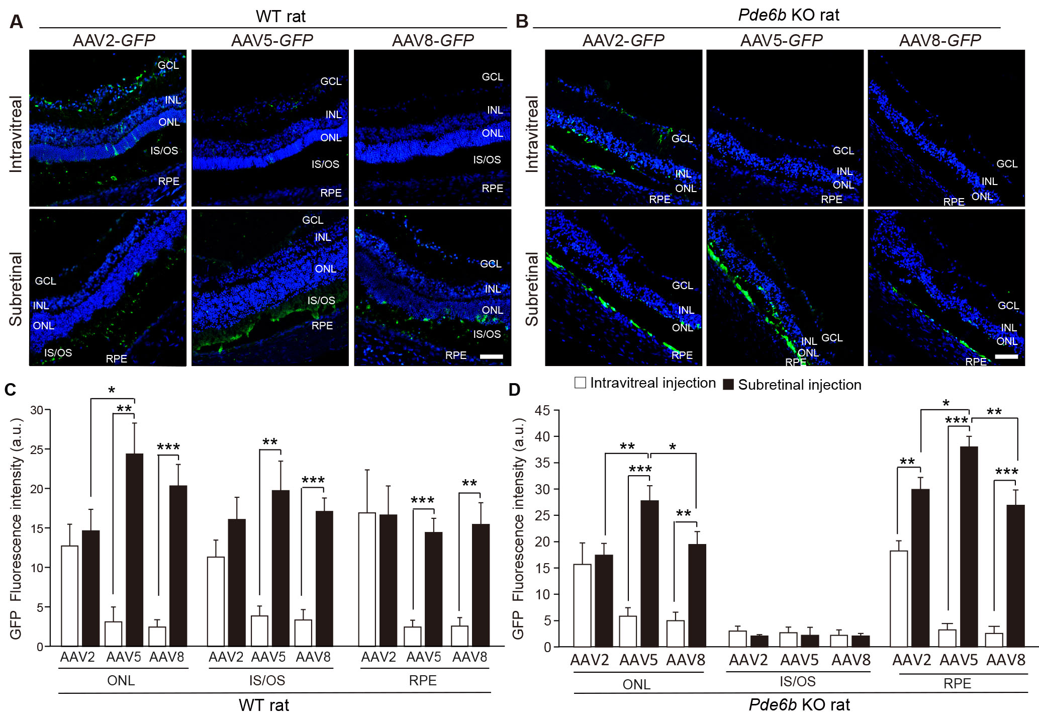
 Figure 2 of
Kim, Mol Vis 2025; 31:127-141.
Figure 2 of
Kim, Mol Vis 2025; 31:127-141.