Figure 1. Noninvasive in vivo green fluorescence protein imaging of 2, 5, and 8 adeno-associated virus serotypes based on the delivery
route in Pde6b knockout rats. A: Schematic diagram of the 8 adeno-associated virus (AAV)-GFP construct. B: Experimental scheme. C–T: Representative fundus photographs showing green fluorescence protein (GFP) expression in live Pde6b knockout rats with intravitreal injection (C–K) or with subretinal injection (L–T) of AAV2-GFP, AAV5-GFP and AAV8-GFP vectors at two, four, and six weeks. U: Quantification of GFP fluorescence intensity from fundus fluorescent photographs. a.u., arbitrary units. n=4 for each group.
Mean fluorescence values were measured over the circular imaging range.
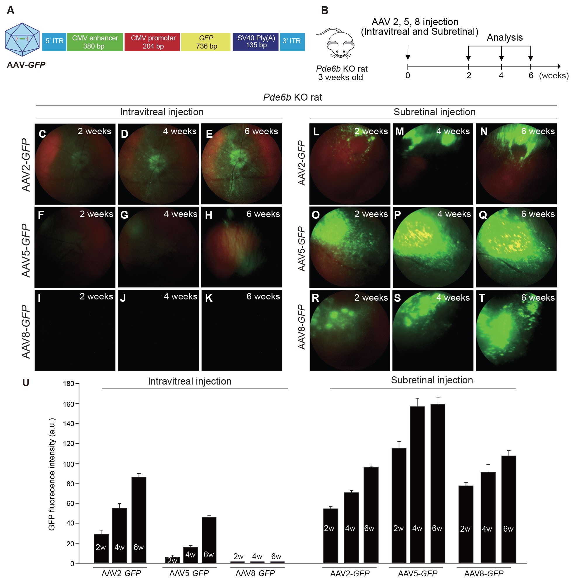
 Figure 1 of
Kim, Mol Vis 2025; 31:127-141.
Figure 1 of
Kim, Mol Vis 2025; 31:127-141.